


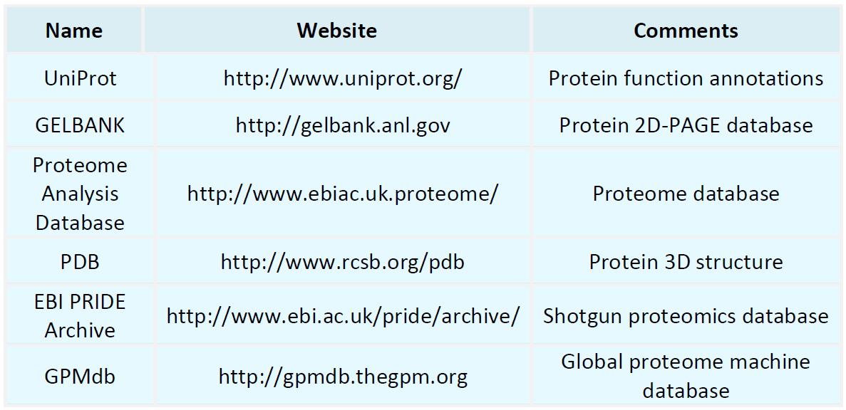
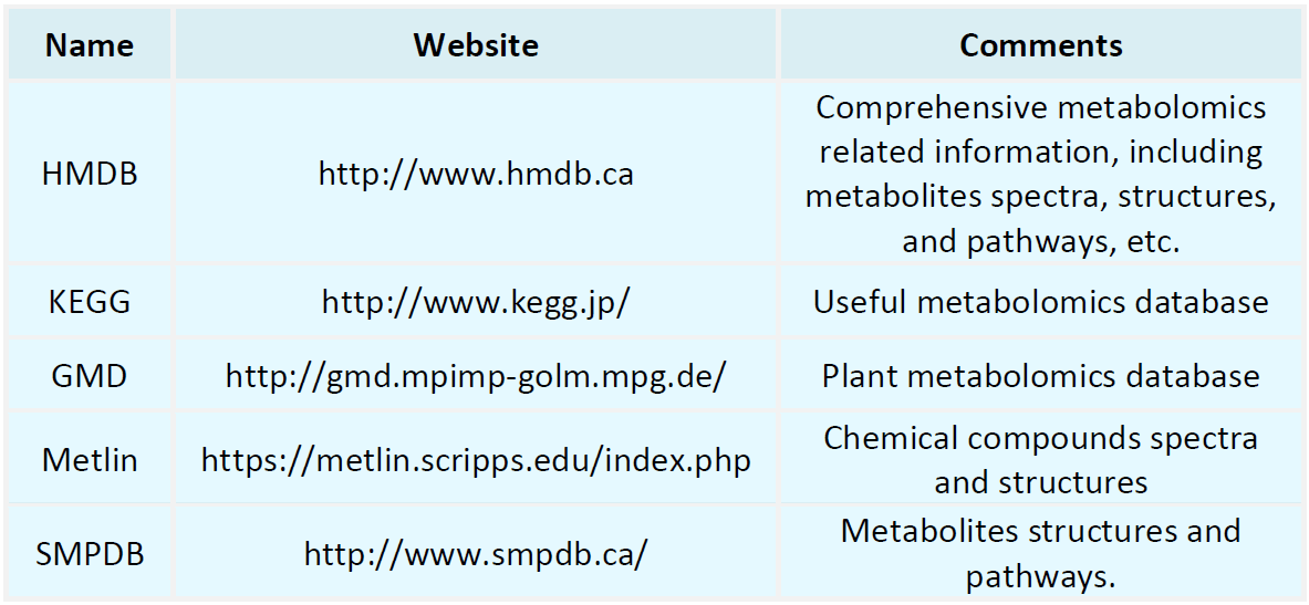
Resources
Proteomics Databases
Metabolomics Databases
-
• Mechanism of Protein Immunoblotting and Electrotransfer
Protein immunoblotting (Western blotting) is a widely used experimental technique in molecular biology, cell biology, and biochemistry research. This technique combines electrophoresis, membrane transfer, and immunodetection methods to detect the presence and quantify specific proteins. This article provides a detailed explanation of the working principle of protein immunoblotting, with a particular focus on the mechanism of electrotransfer.
-
• Application of Protein Immunoblotting and Electrotransfer
Protein immunoblotting (Western blotting) and electrotransfer are indispensable techniques in biological research. They are extensively used for protein identification, expression level analysis, and interaction studies. Due to their precision and sensitivity, these techniques are invaluable across various research fields, including medicine, biochemistry, molecular biology, and drug development. This article explores the applications of protein immunoblotting and electrotransfer.
-
• Workflow of Protein Immunoblotting and Electrotransfer
Protein immunoblotting, commonly known as Western blotting, is a widely utilized experimental technique designed to detect and analyze the presence, expression levels, and molecular weight of specific proteins. The methodology encompasses six primary steps: protein extraction, protein electrophoresis, protein transfer, blocking, antibody incubation, and signal detection. This article provides an in-depth overview of each step within this workflow.
-
• Advantages and Disadvantages of Protein Immunoblotting and Elect
Protein immunoblotting and electrophoretic transfer are essential tools in molecular biology research, widely used for the separation, identification, and analysis of proteins. This article will explore the principles, applications, and advantages and disadvantages of these two techniques in detail.
-
• Principle of Protein Immunoblotting and Electrotransfer
Protein immunoblotting, commonly known as Western blot, is a widely utilized technique in biological research for detecting specific proteins and assessing their expression levels. This method combines gel electrophoresis to separate proteins with immunodetection techniques, based on the specific recognition of target proteins by antibodies.
-
• Workflow of 2D Gel Electrophoresis
Two-Dimensional Gel Electrophoresis (2-DE) is a robust technique widely employed in proteomics to separate proteins based on their isoelectric point (pI) and molecular weight. This article provides a detailed workflow of the 2-DE process.
-
• Principle of 2D Gel Electrophoresis
Two-Dimensional Gel Electrophoresis (2-DE) is a high-resolution protein separation technique widely used in proteomics research. This method combines isoelectric focusing (IEF) and SDS-polyacrylamide gel electrophoresis (SDS-PAGE), enabling the efficient separation of complex protein mixtures based on their isoelectric points (pI) and molecular weights (MW).
-
• Workflow of 1D SDS-PAGE and IEF
Protein separation is a crucial step in molecular biology research. Two commonly used protein separation techniques are one-dimensional SDS-PAGE (Sodium Dodecyl Sulfate-Polyacrylamide Gel Electrophoresis) and Isoelectric Focusing (IEF). These techniques separate proteins based on their different characteristics, utilizing molecular weight and isoelectric point (pI) differences, respectively. This article will detail the workflows of 1D SDS-PAGE and IEF.
-
• Principle of 1D SDS-PAGE and IEF
One-dimensional sodium dodecyl sulfate-polyacrylamide gel electrophoresis (1D SDS-PAGE) and isoelectric focusing electrophoresis (IEF) are widely used protein separation techniques in biological research. These methods exploit differences in the electrophoretic behavior of proteins under various conditions to achieve separation and analysis.
-
• Circular Dichroism Spectrum of Proteins
Circular dichroism (CD) spectroscopy is a commonly used spectral technique for studying the secondary structure of biological macromolecules such as proteins and nucleic acids. This technique is based on the differential absorption of left- and right-handed circularly polarized light by chiral centers in molecules, such as α-helices and β-sheets in proteins.
How to order?

