






How Differential Exosomal Proteins Are Identified and Interpreted
In quantitative exosome proteomics, a differential protein is supported by a measurable abundance difference between predefined groups, not by fold change or presence-versus-absence alone. Reliable differential analysis also requires statistical support, consistent behavior across biological replicates, and sufficient quantitative coverage across the samples being compared. Differential protein analysis is one stage of the broader workflow described in Exosome Proteomics: A Practical Guide for Protein Analysis Studies. This article focuses on how to determine whether quantitative differences are sufficiently supported for biological interpretation and follow-up research.
Researchers planning an exosome proteomics study can contact MtoZ Biolabs to discuss sample grouping, biological replicates, quantitative comparison goals, and analysis requirements.
What Is a Differential Exosomal Protein?
A differential exosomal protein is a protein whose measured abundance differs between predefined sample groups with sufficient quantitative support for that comparison. The groups may represent disease and control samples, treated and untreated conditions, genotypes, time points, or another biological contrast established before analysis.
This is different from comparing two protein identification lists. A protein detected in one group but not another requires separate evaluation because the pattern may reflect either a biological difference or incomplete quantitative coverage. Whether the result supports differential abundance should therefore be determined from the quantitative data rather than detection status alone.
The term “exosomal protein” also refers here to a protein measured in the analyzed exosome preparation. Routine isolation can recover other extracellular vesicles or non-vesicular material, so differential abundance does not by itself establish that a changing protein is exclusively exosome-derived. When the unresolved question concerns peptide-level identification evidence rather than quantitative group differences, LC-MS/MS-Based Exosome Protein Profiling provides the more relevant framework.
How Are Differential Exosomal Proteins Identified?
Differential analysis begins with a quantitative protein matrix containing protein measurements for individual samples assigned to predefined groups. The analysis then asks whether the matrix provides enough evidence to distinguish a group-associated abundance change from ordinary variation or incomplete measurement.
1. Quantitative Change Defines What Is Being Compared
Abundance change establishes the direction and apparent magnitude of the difference between groups. Fold change is useful for describing that effect, but it does not determine whether the effect is sufficiently supported. Two proteins with similar fold changes can be supported by very different quantitative data, so a large numerical change should not be prioritized automatically. Candidate prioritization should consider sample-level measurements rather than relying on the group-average fold change alone.
2. Statistical Support Relates the Group Difference to Sample Variation
Statistical evidence evaluates the observed group separation in relation to variation within the groups. When biological replicate measurements are relatively concentrated, a between-group shift can be distinguished more clearly from background variation; when replicate values are widely dispersed, the same difference between group averages provides weaker evidence for a stable group-associated change. No universal fold-change or statistical threshold is appropriate for every exosome proteomics dataset, so screening criteria should be interpreted in the context of the study design and the actual distribution of measurements rather than treated as sufficient evidence on their own.
3. Biological Replicates Show Whether the Change Represents the Group
Biological replicates reveal whether a measured change recurs across independent samples or is dominated by isolated observations. A protein whose abundance shifts in the same direction across most replicates provides stronger evidence for a group-associated difference than one whose apparent change depends largely on one extreme sample. A candidate driven by a small number of samples may still be biologically relevant, but it should not be prioritized simply because its average fold change is large; the sample-level pattern should first support the interpretation that the change represents the predefined biological groups.
4. Data Completeness Determines Whether the Comparison Is Direct
Quantitative completeness determines how directly abundance can be compared across groups. A protein measured across most samples in both groups provides a more direct basis for estimating a group difference than one represented by sparse or strongly asymmetric measurements. Stable measurements in one group combined with frequent missing values in another may still indicate a biologically meaningful change, but the evidence contains a stronger detection component and should not be interpreted in the same way as a consistently quantified abundance shift. When missingness becomes a major feature of the result, the quantitative matrix should be reviewed to determine whether the protein belongs in the main differential set, requires separate interpretation, or needs additional evidence.
After quantitative change, statistical support, biological replicate behavior, and data completeness have been evaluated together, the resulting differential protein list can be assessed for biological consistency and interpretation.
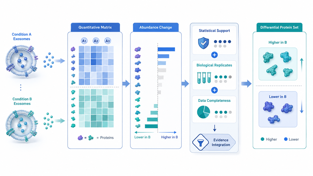
Figure 1. Evidence Integration from Quantitative Matrix to Differential Protein Set
How Should Differential Protein Results Be Evaluated?
Once the differential protein set has been generated, result evaluation moves from individual screening criteria to the structure of the dataset. The central question is whether the selected proteins collectively represent the intended biological comparison strongly enough to support downstream interpretation.
1. Does the Differential Set Reflect the Intended Comparison?
A useful differential result should describe the predefined groups more clearly than it describes a few unusual samples. Sample-level abundance patterns can reveal whether the selected proteins remain broadly aligned with the biological grouping or whether a substantial part of the result is concentrated in atypical observations. If the final list contains many proteins but only a small subset shows stable patterns across samples, the total differential count is less informative than the composition of the list, and downstream interpretation should focus first on the proteins whose quantitative behavior is compatible with the comparison the experiment was designed to test.
2. Which Results Are Ready for Biological Interpretation?
Differential proteins do not all carry the same interpretive weight after screening. The practical question is which results can move directly into biological interpretation and which require closer review of the underlying quantitative data. The result patterns below lead to different next decisions.
|
Result Pattern |
What It Means |
Practical Next Step |
|
Differential proteins show a clear sample-level pattern aligned with the predefined groups |
Stronger basis for group-level interpretation |
Proceed to biological pattern analysis |
|
The list is dominated by proteins supported by only a small subset of samples |
The nominal differential set may overstate stable group-associated evidence |
Review sample-level measurements before prioritizing candidates |
|
Many proteins show extensive or asymmetric missing measurements |
Part of the result may reflect detection differences rather than direct abundance comparison |
Separate well-quantified changes from detection-dominated signals |
|
Multiple proteins show coordinated condition-associated changes |
The dataset provides a basis for functional or pathway-level interpretation |
Examine whether the changes converge on related biological processes |
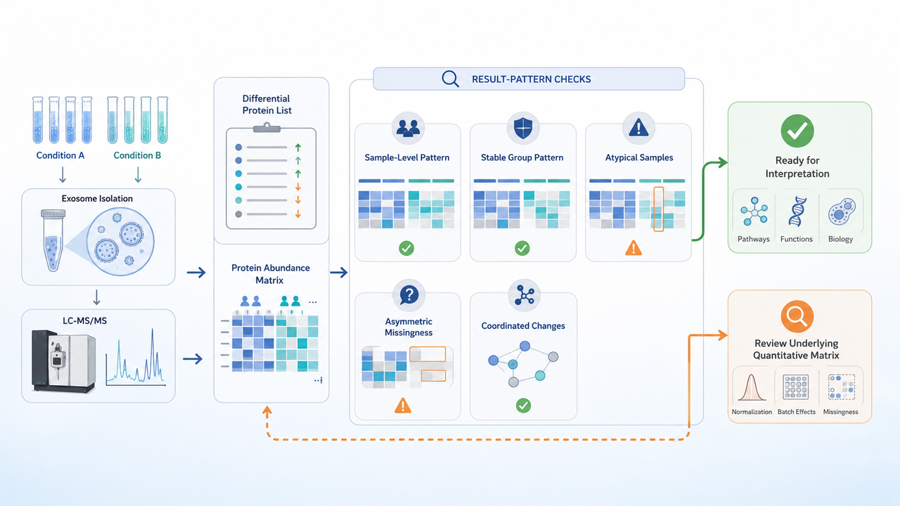
Figure 2. Result-Pattern Checks Before Biological Interpretation
When a differential list is dominated by unstable sample patterns, extensive missing measurements, or changes supported by only a small number of observations, adding more functional interpretation immediately may not resolve the uncertainty. The quantitative matrix and sample-level distributions should first be reviewed to determine which results are sufficiently supported for downstream analysis. If your differential exosome proteomics results contain extensive missing values, large replicate variability, or an inconsistent candidate list, contact MtoZ Biolabs to discuss the study design, quantitative data, comparison groups, and biological objective.
Biological Interpretation of Differential Protein Patterns
Once the quantitative result has been narrowed to changes that are sufficiently supported for interpretation, the next step is to determine whether those changes point to isolated protein signals or a broader biological pattern.
1. Individual Protein Changes Define Specific Research Leads
An individual differential protein can provide a focused research lead when its known function is relevant to the original biological comparison. At this stage, the question shifts from whether the protein is differential to what biological question the change raises. A protein does not need to belong to a large enriched pathway to be worth following; a well-supported change may be informative because it relates directly to a known mechanism, phenotype, molecular process, or prior experimental observation. Its value therefore depends on how clearly the quantitative result connects to the study question and what specific hypothesis that connection suggests.
2. Coordinated Protein Changes Provide Biological Context
When several differential proteins are linked to related molecular functions or processes, the result becomes easier to interpret at a systems level. In exosome preparations, coordinated changes may indicate that the measured protein composition shifts with the experimental condition around a common biological theme. The value of this pattern lies in convergence rather than in the number of proteins alone: related changes can support a more specific biological hypothesis and help determine which candidates or processes deserve follow-up, while isolated and coordinated signals may reasonably lead to different experimental questions.
3. Functional Annotation and Pathway Enrichment Refine the Biological Question
Functional annotation and pathway enrichment organize the differential protein set according to known biological functions and pathways, helping identify where the changing proteins converge and whether those patterns fit the original experimental question. An enriched pathway should be interpreted as an association within the measured protein pattern rather than as direct evidence that the pathway is activated, inhibited, or mechanistically responsible for the phenotype. The value of the analysis is therefore to narrow the biological question and provide a rationale for targeted follow-up, while the subsequent experiment determines whether the proposed relationship actually holds.
How Can the Interpretability of Differential Exosome Proteomics Results Be Strengthened?
Once the quantitative and biological patterns have been defined, the remaining task is to ensure that the next conclusion or experiment matches the evidence already obtained.
1. Keep Exosome Preparation Context Attached to the Result
Differential protein interpretation should remain connected to how the exosome preparations were generated. If comparison groups differ systematically in isolation, purification, storage, or upstream handling, part of the measured protein difference may reflect preparation history rather than the biological condition being tested. This does not mean that every project requires identical handling under all circumstances, but systematic preparation differences need to be separated from biological differences before the quantitative pattern is given a strong biological interpretation.
When preparation history becomes a major source of uncertainty, How Exosome Isolation and Purification Affect Proteomics Results addresses how recovery, background, and preparation composition can alter the measured proteome.
2. Match the Next Experiment to the Question the Result Raises
The next step should be determined by the specific uncertainty left by the differential result rather than by applying the same follow-up strategy to every candidate.
● Confirm whether selected protein changes are reproducible
If the main uncertainty is whether selected proteins consistently differ between groups, the next step should strengthen quantitative evidence around those targets. This is most relevant when candidates have already been identified but their abundance differences still require more focused confirmation.
● Test whether a candidate contributes to the biological effect
If the research question moves from association to function, additional differential profiling is unlikely to resolve it. Functional experiments are needed to determine whether the candidate contributes directly to the observed phenotype or biological response.
● Evaluate whether a candidate has diagnostic or prognostic value
If the objective shifts from discovery to biomarker performance, the study design must move beyond the original differential dataset. Independent samples and validation designed around diagnostic or prognostic performance are required before that type of conclusion can be supported.
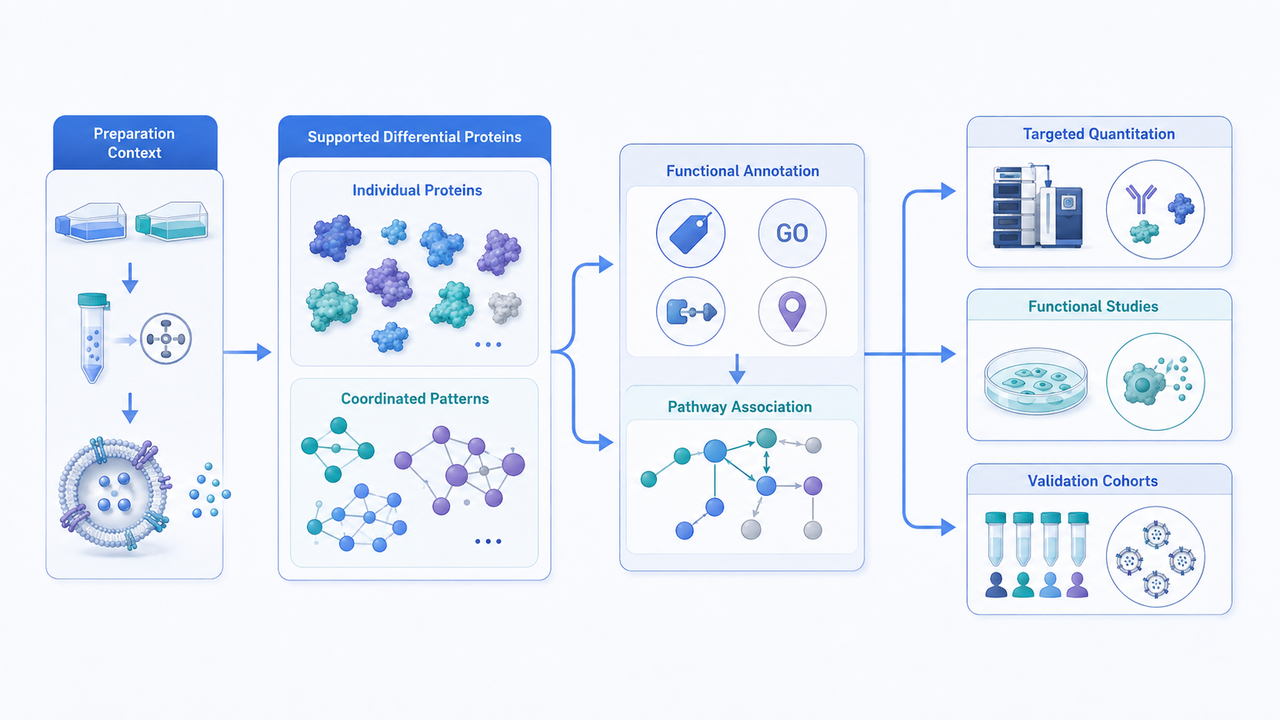
Figure 3. From Differential Protein Patterns to Follow-Up Study Design
Conclusion
Researchers who already have defined sample groups, biological replicates, and a quantitative comparison objective can review the MtoZ Biolabs Exosome Protein Analysis Service for project-specific evaluation. When differential results are difficult to interpret because of group variability, incomplete quantification, preparation differences, or uncertainty about the next analytical step, the study design, quantitative data structure, and intended biological question can be reviewed together to determine the most appropriate analysis and interpretation path.
How to order?




