






From Differential Proteins to Mitochondrial Pathways: How to Interpret Proteomics Data
A mitochondrial proteomics experiment can generate hundreds or even thousands of quantified proteins, but a differential protein list does not automatically explain the biology. This is where many studies become difficult to interpret. A volcano plot shows which proteins increase or decrease. A heatmap reveals patterns across samples. Enrichment analysis may return dozens of statistically significant terms. Yet none of these outputs alone answers the most important question: What do these protein changes mean for mitochondrial biology?
The challenge is to move from statistical differences to a biologically defensible interpretation without overemphasizing individual proteins, enrichment P values, or predicted networks. A practical interpretation workflow follows five steps: Differential Protein Screening, Functional Enrichment, Mitochondrial Pathway Prioritization, Network Context and Hypothesis Building, Complementary Evidence and Targeted Validation
The purpose of this workflow is not to force every dataset into a mitochondrial story. It is to determine whether the results show coherent mitochondrial remodeling and which proteins or pathways deserve further investigation.
Step 1: Prioritize Differential Proteins Beyond P Values
Differential analysis is usually the first step after quantitative proteomics data have passed basic quality control. Common parameters include fold change, P value, adjusted P value or false discovery rate, replicate number, and within-group variation. These metrics are useful, but none should be interpreted alone.
A protein with a large fold change but poor consistency across biological replicates may be less convincing than a protein with a smaller but reproducible change. Likewise, a rigid fold-change cutoff may remove mitochondrial proteins that change only modestly but participate in a coordinated pathway-level response.
Treat Statistical Thresholds as Filters, Not Biological Rules
A cutoff such as FC >1.5 and P <0.05 may be useful for organizing a dataset, but it should not become a universal definition of biological relevance.
For mitochondrial studies, candidate prioritization should also consider:
- replicate consistency;
- data completeness and missing values;
- protein abundance;
- mitochondrial localization evidence;
- pathway membership;
- coordinated changes among related proteins.
This is especially important for lower-abundance regulatory proteins and components of multiprotein mitochondrial complexes.
Review the Overall Data Structure First
Different visualization tools answer different questions. Volcano plots summarize effect size and statistical significance. Heatmaps help determine whether selected proteins show consistent patterns across samples. PCA can reveal overall sample relationships, replicate clustering, and potential outliers.
Missing-value handling also deserves attention. Imputation can influence downstream differential analysis, particularly when missingness is associated with low protein abundance.
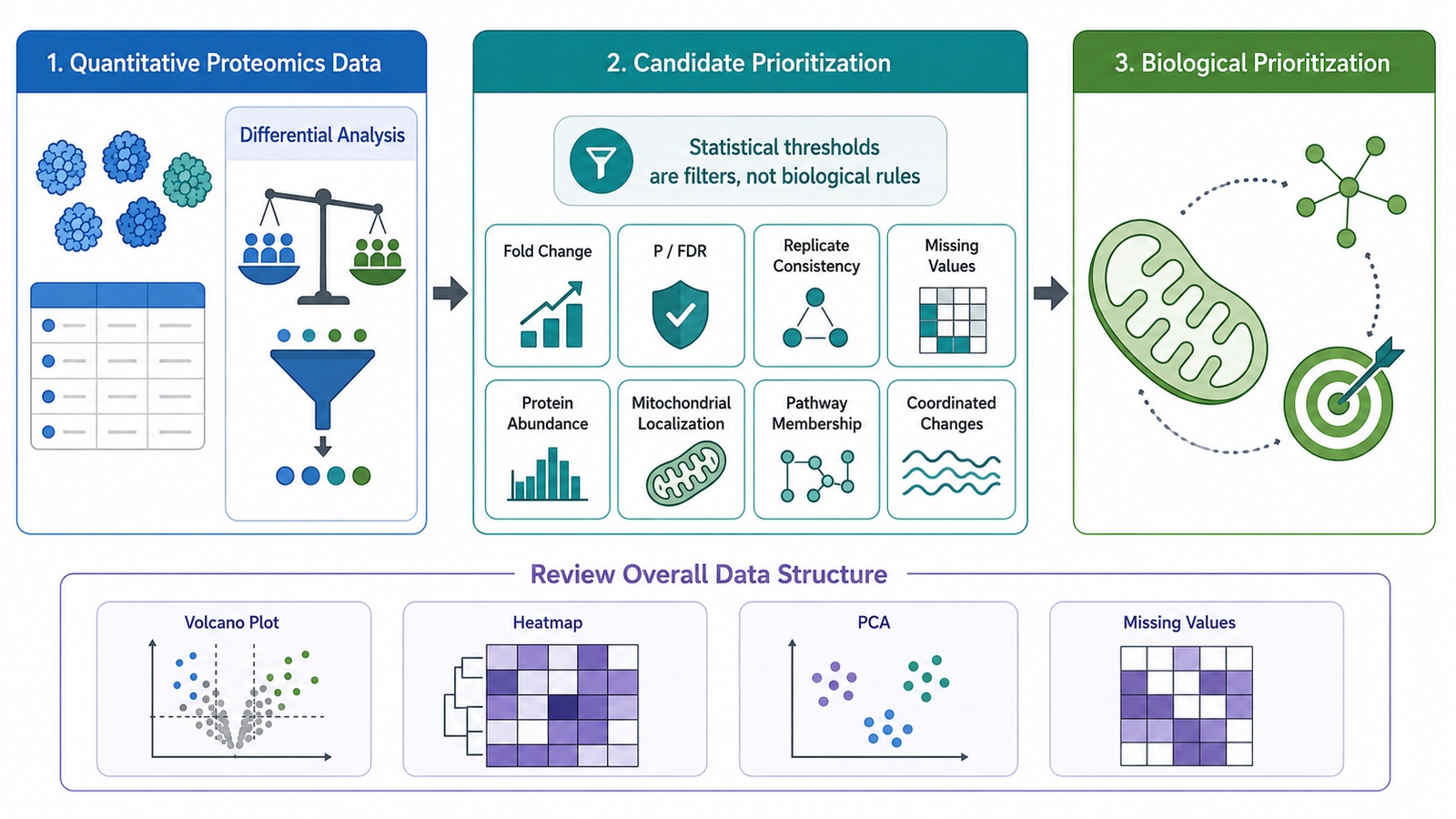
Figure 1. From Quantified Proteins to Biological Prioritization.
Reliable interpretation begins before differential analysis. If you are planning a mitochondrial proteomics study, MtoZ Biolabs can support the workflow from mitochondrial sample preparation and quantitative proteomics to downstream data analysis and biological interpretation. Contact us to discuss your sample type, study design, and comparison strategy.
Step 2: Use Functional Enrichment to Identify Biological Themes
Once a differential protein set has been defined, functional enrichment helps answer a broader question:
Which biological processes are represented more strongly than expected in the protein data?
At this stage, the objective is to identify major biological themes rather than immediately assign every result to a mitochondrial mechanism.
Choose the Enrichment Approach According to the Question
Over-Representation Analysis (ORA) tests whether a predefined differential protein list contains more members of a pathway or functional category than expected.
Its main limitation is that the result depends on the cutoff used to define the differential protein set.
Gene Set Enrichment Analysis (GSEA) uses a ranked list rather than relying only on proteins that pass a strict threshold. It can therefore identify coordinated pathway-level shifts even when many individual proteins show relatively modest changes.
ssGSEA or GSVA may be useful when pathway-level scores are needed at the individual-sample level.
Use Annotation Databases to Organize, Not Multiply, Results
Common resources include:
- GO;
- KEGG;
- Reactome;
- WikiPathways.
The purpose is not to report every significant term. Closely related or redundant terms should be grouped into broader biological themes.
Interpretation should consider more than enrichment P value. Useful questions include:
- How many measured proteins contribute to the term?
- Are the protein changes directionally consistent?
- Are important pathway components represented?
- Does the pathway fit the experimental question?
- Are several significant terms describing essentially the same biological process?
If mitochondrial-related themes emerge at this stage, the next task is not simply to report them. The next task is to determine which differential proteins have direct mitochondrial evidence and how they map to specific mitochondrial systems.
Step 3: Apply a Mitochondrial-Specific Interpretation Layer
This is the step that turns general proteomics interpretation into mitochondrial-focused analysis.
A useful approach is to divide relevant differential proteins into three groups.
1. Proteins With Established Mitochondrial Localization
These proteins have evidence supporting localization within or association with mitochondria.
Resources such as MitoCarta, MitoMiner, IMPI, UniProt, and subcellular localization information from the Human Protein Atlas can support this classification.
2. Proteins Associated With Mitochondrial Pathways
Some proteins participate in pathways that influence mitochondrial biology but are not necessarily exclusive mitochondrial residents.
These proteins may still be highly relevant to the biological interpretation, but their localization should be described accurately.
3. Upstream Regulators of Mitochondrial Biology
A cytosolic or nuclear regulatory protein may affect mitochondrial metabolism, dynamics, stress responses, or biogenesis without itself being a mitochondrial protein.
Keeping this group separate prevents a common interpretation error: calling every protein involved in a mitochondrial pathway a “mitochondrial protein.”
4. Map the Filtered Proteins to Mitochondrial Systems
After this classification, proteins can be organized into specific mitochondrial processes.
For OXPHOS, examine whether changes occur across several components of electron transport chain complexes rather than highlighting one subunit in isolation.
For the TCA cycle, look for coordinated remodeling across multiple metabolic enzymes.
For mitochondrial dynamics, proteins associated with fusion and fission can be interpreted as a functional group.
Mitophagy, protein import, metabolite transport, and apoptosis-related mitochondrial signaling can be evaluated in the same way.
The key question is:
Do multiple proteins support a coherent change in the same mitochondrial process?
That is usually more informative than simply selecting the proteins with the largest fold changes.
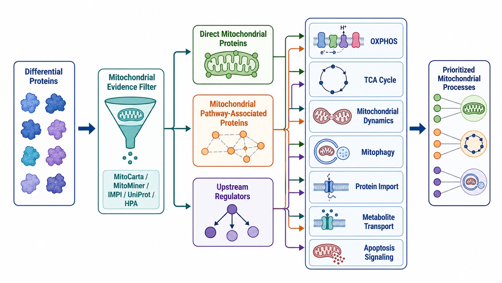
Figure 2. Mitochondrial-Specific Filtering of Differential Proteins.
Step 4: Build Mechanistic Hypotheses Without Overinterpreting Networks
Once mitochondrial pathways have been prioritized, network and regulatory analyses can help organize the findings into testable hypotheses.
They should not be treated as proof of mechanism.
Use Protein Networks to Add Context
STRING can provide known and predicted functional associations among proteins. Cytoscape can be used to visualize these relationships, while approaches such as MCODE can identify densely connected network modules.
These analyses can help prioritize candidates that sit within a coordinated mitochondrial module.
However, a highly connected protein is not automatically the causal driver of the phenotype.
A stronger prioritization framework is:
Differential Change + Mitochondrial Evidence + Pathway Position + Network Context
Network analysis should therefore help answer which candidates deserve follow-up, not claim which protein caused the entire response.
Treat Upstream Regulatory Analysis as Hypothesis Generating
When appropriate data are available, regulatory enrichment can provide additional context.
For example, transcription-factor enrichment may suggest regulatory programs associated with a differential protein set. ChIP-based target resources can also generate hypotheses about transcriptional control.
Kinase-substrate enrichment is more informative when regulated phosphorylation sites are available from phosphoproteomics, rather than from total protein abundance data alone.
These analyses predict possible regulatory relationships. They do not directly demonstrate transcription-factor activity, kinase activity, or causal regulation.
Compare Proteomic Patterns With Independent Phenotypes
If independent functional data are available, they can strengthen interpretation.
For example, coordinated changes in OXPHOS proteins may be considered together with separately measured mitochondrial respiration, ATP-related readouts, membrane potential, ROS, mtDNA-related measurements, or imaging phenotypes.
These are complementary evidence layers.
Proteomics itself does not directly measure these mitochondrial functional endpoints.
Step 5: Add Complementary Evidence and Targeted Validation
After the mitochondrial pathway and candidate list have been narrowed, the next experiment should address the largest remaining uncertainty.
Proteomics Plus Metabolomics
Metabolomics is particularly useful when the proteomics result points toward the TCA cycle, fatty-acid oxidation, amino-acid metabolism, or another metabolic pathway.
Proteomics may identify coordinated changes in enzymes or transporters, while metabolomics can determine whether related metabolites also change.
This provides complementary pathway-level evidence, although metabolite abundance alone does not demonstrate metabolic flux.
Proteomics Plus Transcriptomics
Transcriptomics can determine whether protein abundance changes are accompanied by corresponding RNA changes.
Agreement between RNA and protein may support a coordinated expression program. Discordance can also be informative and may point toward post-transcriptional regulation, translation, protein stability, or turnover.
Use Targeted Validation for the Most Important Claims
Western blot or targeted PRM can provide follow-up measurements for selected candidate proteins.
The purpose is not to repeat the entire discovery dataset. Validation should focus on proteins that are central to the proposed pathway or hypothesis.
Functional follow-up should also match the biological claim. If a study proposes altered mitochondrial respiration, membrane potential, ROS, or morphology, the corresponding functional assay should be planned independently.
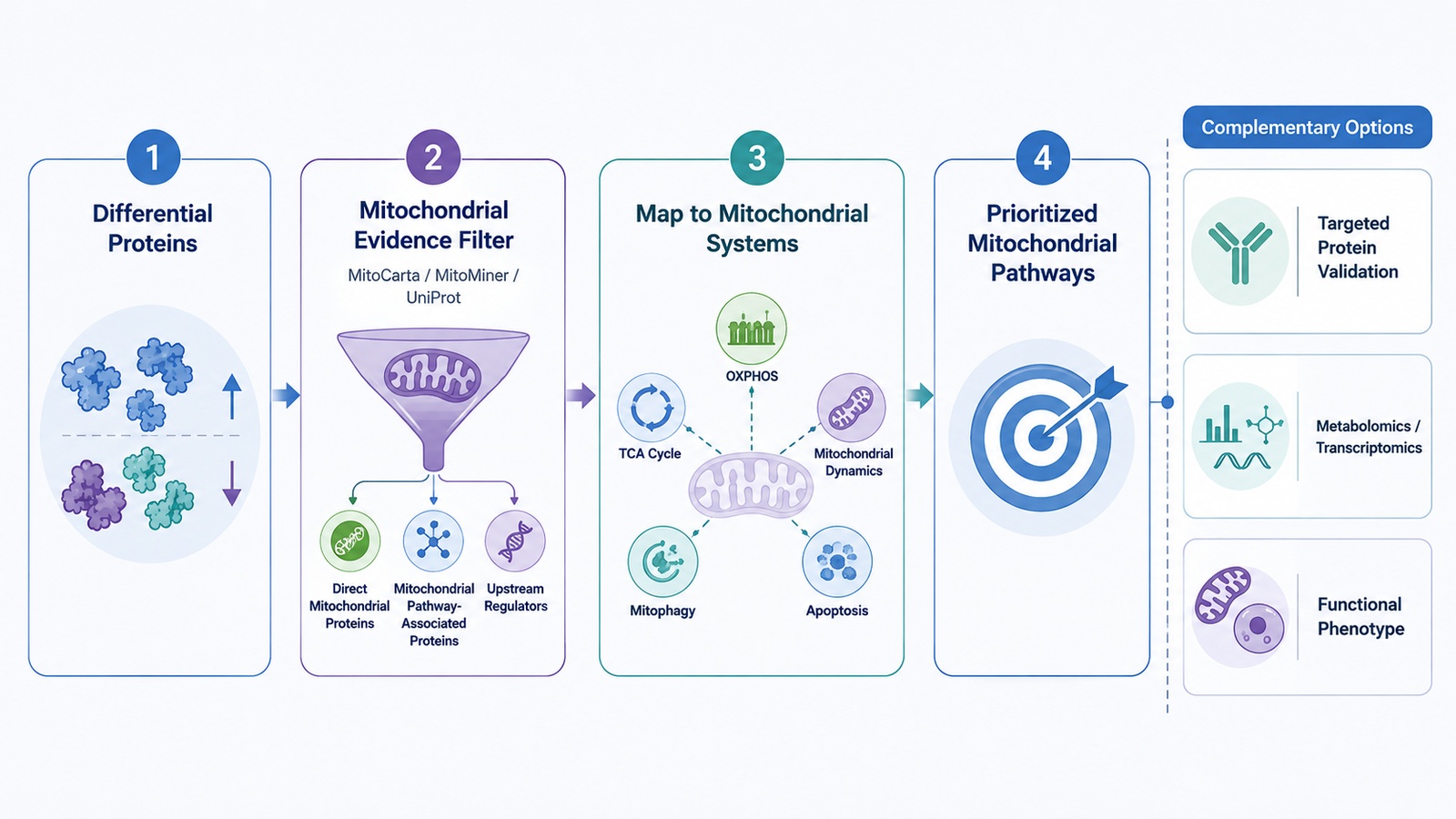
Figure 3. From Differential Proteins to Prioritized Mitochondrial Pathways.
Related Services
Mitochondrial Proteomics Service
Subcellular Proteomics Service
Subcellular Structure and Organelle Proteomics Service
Five Common Mistakes in Mitochondrial Proteomics Interpretation
1. Ranking Pathways by P Value Alone
Statistical enrichment should be interpreted together with pathway coverage, protein directionality, and biological relevance.
2. Calling Every Mitochondrial Regulator a Mitochondrial Protein
Subcellular localization evidence should be reviewed before assigning mitochondrial identity.
3. Reporting Differential Proteins Without Pathway Context
Individual proteins become more informative when interpreted together with related pathway components and protein complexes.
4. Applying Fold-Change Thresholds Mechanically
Moderate but coordinated protein changes may be biologically relevant even when individual proteins fall below an arbitrary cutoff.
5. Treating Protein Abundance as Mitochondrial Function
Protein abundance does not directly measure enzyme activity, metabolite levels, respiration, membrane potential, ROS, or other functional states.
Conclusion
The value of mitochondrial proteomics extends beyond differential protein lists. Meaningful interpretation should progress from differential protein screening to functional enrichment, mitochondrial-specific filtering, pathway prioritization, network context, hypothesis building, and targeted follow-up. Each step should refine the biological question, while clearly distinguishing mitochondrial-localized proteins, mitochondrial pathway-associated proteins, and upstream regulators to support more precise and defensible conclusions.
MtoZ Biolabs supports mitochondrial protein identification, quantitative proteomics, PTM analysis, mitochondrial protein annotation, pathway analysis, and proteomics-metabolomics integration. If you are planning a mitochondrial proteomics study, you can provide your research question, sample type, comparison design, and current data status for project evaluation.
How to order?




