






Exosome Proteomics Analysis Service for EV Protein Profiling and Comparative Research
Peptide signals acquired from an extracellular vesicle preparation do not all support the same type of conclusion. Protein identification establishes which proteins or protein groups are represented by the detected peptide evidence. Quantitative profiling evaluates relative abundance patterns across samples, while differential analysis identifies proteins associated with a predefined comparison.
LC-MS/MS exosome proteomics connects these evidence levels within a single analytical framework. EV protein profiling can describe the protein composition of an EV-enriched preparation, compare experimental groups, and generate candidate proteins for further investigation. The interpretation remains dependent on EV preparation quality, study design, and the distinction between discovery-stage results and independently validated conclusions.
Analytical Scope of Exosome Proteomics
1. EV Protein Identification
EV-associated proteins are commonly analyzed through bottom-up proteomics. Proteins are extracted from the EV preparation, digested into peptides, separated by liquid chromatography, and measured by tandem mass spectrometry. Peptide-spectrum evidence is then matched against a sequence database and assembled into protein or protein-group identifications.
Protein identification establishes that compatible peptide evidence was detected in the analyzed material. It does not determine whether every protein was located inside a vesicle, associated with the vesicle surface, or retained as co-isolated material. The phrase EV cargo profiling is therefore most accurate when the preparation and supporting evidence justify a cargo-focused interpretation. In broader discovery studies, EV-associated protein profiling is often the more precise description.
Identification also differs from quantification. A protein may be detected without showing a measurable difference between groups, while a protein absent from a final differential list may still be consistently present in the analyzed samples.
2. Quantitative EV Protein Profiling
Quantitative proteomics compares relative peptide or protein abundance across predefined samples or biological groups. The resulting abundance matrix supports evaluation of shared proteins, group-associated patterns, sample relationships, and the direction and magnitude of protein changes.
Relative quantification may use a Label-Free design or isobaric labeling with TMT or iTRAQ. Label-Free analysis measures samples separately and depends strongly on consistent preparation and acquisition. Isobaric labeling combines labeled samples within a multiplex and requires deliberate channel allocation and batch planning. Data-independent acquisition (DIA) is a mass spectrometric acquisition strategy rather than a labeling category and is frequently used to produce consistent quantitative matrices in Label-Free studies.
The appropriate route depends on sample number, available material, group structure, batch requirements, and the intended comparison. No single strategy is universally preferable for all EV projects.
3. Differential Protein Analysis
Differential protein analysis evaluates whether relative abundance patterns differ between predefined groups. A defensible analysis considers effect size, biological variability, replicate consistency, missing-value patterns, statistical significance, and multiple-testing control rather than relying on fold change alone.
The resulting differential proteins are discovery-stage candidates associated with the current comparison. They do not automatically represent validated biomarkers, causal regulators, confirmed disease mechanisms, or proteins uniquely localized to EVs. Their value lies in narrowing a broad quantitative dataset into a traceable set of proteins for functional interpretation and follow-up evaluation.
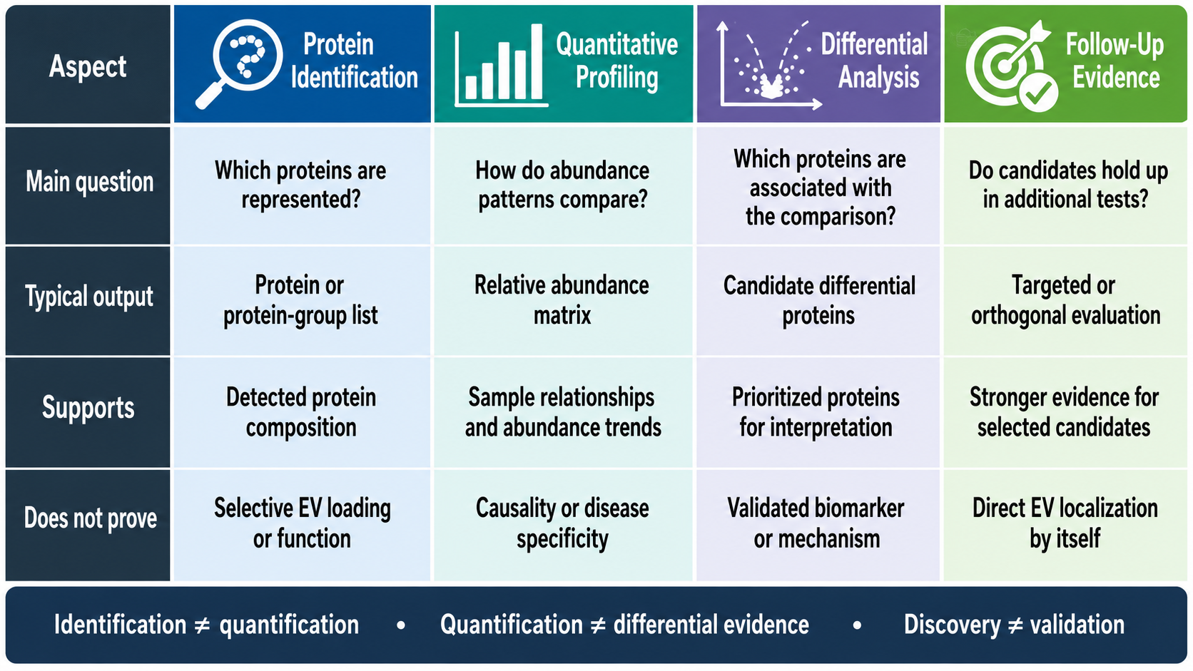
Figure 1. Evidence Levels in EV Proteomics Analysis
Research Projects Suited to EV Proteomics
1. Biological Group Comparisons
EV proteomics can compare protein profiles across disease-related and control groups, distinct phenotypes, experimental models, or other biologically defined cohorts. Such studies may reveal proteins whose relative abundance differs between groups and may identify pathways or protein networks associated with the study condition.
Interpretation depends on the comparability of sample collection, EV preparation, protein processing, and batch allocation. A group-associated protein difference supports an association with the defined comparison but does not independently establish clinical specificity, diagnostic performance, or biological causation.
2. Treatment and Time-Course Studies
Treatment-response studies can examine how EV-associated protein profiles change after pharmacological exposure, environmental perturbation, genetic manipulation, or another controlled intervention. Time-course designs can evaluate whether protein abundance patterns emerge, persist, or return toward baseline across predefined sampling points.
These projects require careful separation of biological time or treatment effects from preparation batches. Collection timing, cell state, EV isolation, sample processing, and LC-MS/MS acquisition should be balanced across groups. A temporal trend represents comparative evidence and still requires additional experiments before it can be interpreted as a functional response mechanism.
3. Source- and Model-Dependent EV Profiles
EV protein profiling can also compare vesicles derived from different cell models, culture conditions, biological fluids, tissues, or experimental systems. Source-dependent studies may describe broad differences in protein composition, whereas comparative quantitative studies require sufficiently matched groups to support relative abundance testing.
Differences between plasma-, urine-, CSF-, or cell culture-derived EV preparations may reflect both biology and source-specific matrix effects. Comparisons across distinct sample types therefore require more cautious interpretation than comparisons performed within a single source under controlled conditions.
From EV Material to LC-MS/MS Data
1. Starting Material and EV Preparation Status
(1) Source Samples and Pre-Isolated EVs
EV proteomics may begin with plasma, serum, urine, cerebrospinal fluid, cell culture-conditioned medium, or another appropriate source sample. These materials generally require EV separation, enrichment, or purification before protein preparation. The selected approach influences protein recovery, non-vesicular background, and the interpretation of the resulting profile.
Pre-isolated EVs can enter the workflow at a later stage, but their suitability depends on the original source, separation method, purity, integrity, buffer composition, storage history, and available quality-control information. The label “isolated EVs” alone does not establish that the preparation is ready for LC-MS/MS.
(2) EV Lysates and Prepared Peptides
EV protein lysates represent a later analytical starting point. Their compatibility depends on protein recovery, lysis chemistry, detergent content, salt concentration, and other additives that may affect digestion or mass spectrometric analysis.
Prepared peptides bypass EV separation, protein extraction, and digestion. Their assessment focuses on peptide amount, digestion quality, contamination, storage history, and LC-MS/MS compatibility. At this stage, limitations introduced during EV preparation or protein recovery are difficult to correct.
2. Protein Preparation and LC-MS/MS Measurement
Proteomic preparation typically includes protein solubilization, reduction, alkylation, enzymatic digestion, and peptide cleanup. These steps convert EV-associated proteins into peptides that are compatible with chromatographic separation and tandem mass spectrometry.
Liquid chromatography reduces sample complexity by separating peptides before ionization. Tandem mass spectrometry then records precursor and fragment-ion information used for peptide identification and quantitative measurement. Data processing connects accepted peptide evidence to protein groups and, in quantitative projects, generates relative abundance values across samples.
Variation in extraction efficiency, digestion completeness, peptide recovery, chromatography, or acquisition may influence both identification and quantification. Consistent processing is therefore particularly important in comparative studies.
3. Factors Affecting Proteome Coverage
The number and types of proteins detected in an EV preparation depend on several interacting factors. EV purity and co-isolated background influence which peptide signals dominate the analysis. Starting material, biological source, species, EV recovery, protein extraction, digestion quality, chromatographic performance, and LC-MS/MS strategy also affect coverage.
Greater sample input does not necessarily compensate for poor purity or incompatible preparation chemistry. Conversely, low-input samples may still produce interpretable data when preparation loss and background are carefully controlled. Proteome depth should therefore be treated as project-dependent rather than represented by a fixed expected protein count.
Interpreting EV Proteomics Results
1. Identification and Quantitative Outputs
A protein identification table records the proteins or protein groups supported by accepted peptide evidence. Depending on the analysis, it may also include peptide counts, sequence coverage, confidence metrics, or other identification-level information.
A quantitative matrix presents relative protein abundance across samples. Quality-control summaries and standard visualizations can be used to examine data distributions, replicate relationships, clustering patterns, and group separation. Differential analysis then identifies proteins associated with the predefined comparison.
These outputs answer related but distinct questions. Identification describes detected protein composition, quantification describes relative abundance, and differential testing evaluates evidence for group-associated changes.
2. Functional Analysis and Candidate Prioritization
Gene Ontology, KEGG pathway, and protein-protein interaction analyses can organize identified or differential proteins into functional categories, pathways, and network relationships. These analyses support biological interpretation but depend on existing annotations, database coverage, and the proteins included in the analysis.
Candidate prioritization may consider effect size, replicate consistency, peptide evidence, data completeness, functional annotation, network position, known cellular distribution, and the study objective. A statistically significant protein is not automatically the most biologically relevant candidate, and database association does not establish experimental function.
3. Discovery Results and Follow-Up Evidence
Differential proteins and prioritized candidates remain discovery-stage results until they are evaluated through additional evidence. PRM-based targeted mass spectrometry can assess selected peptides with a more focused measurement strategy. Western blotting or ELISA may offer orthogonal protein-level evaluation when suitable antibodies, sample material, and assay conditions are available.
Follow-up measurements may support repeated detection or confirmation of a group-associated abundance pattern. Broader conclusions regarding protein function, EV localization, mechanism, clinical relevance, or biomarker performance require independent samples, appropriate controls, and additional experimental validation.
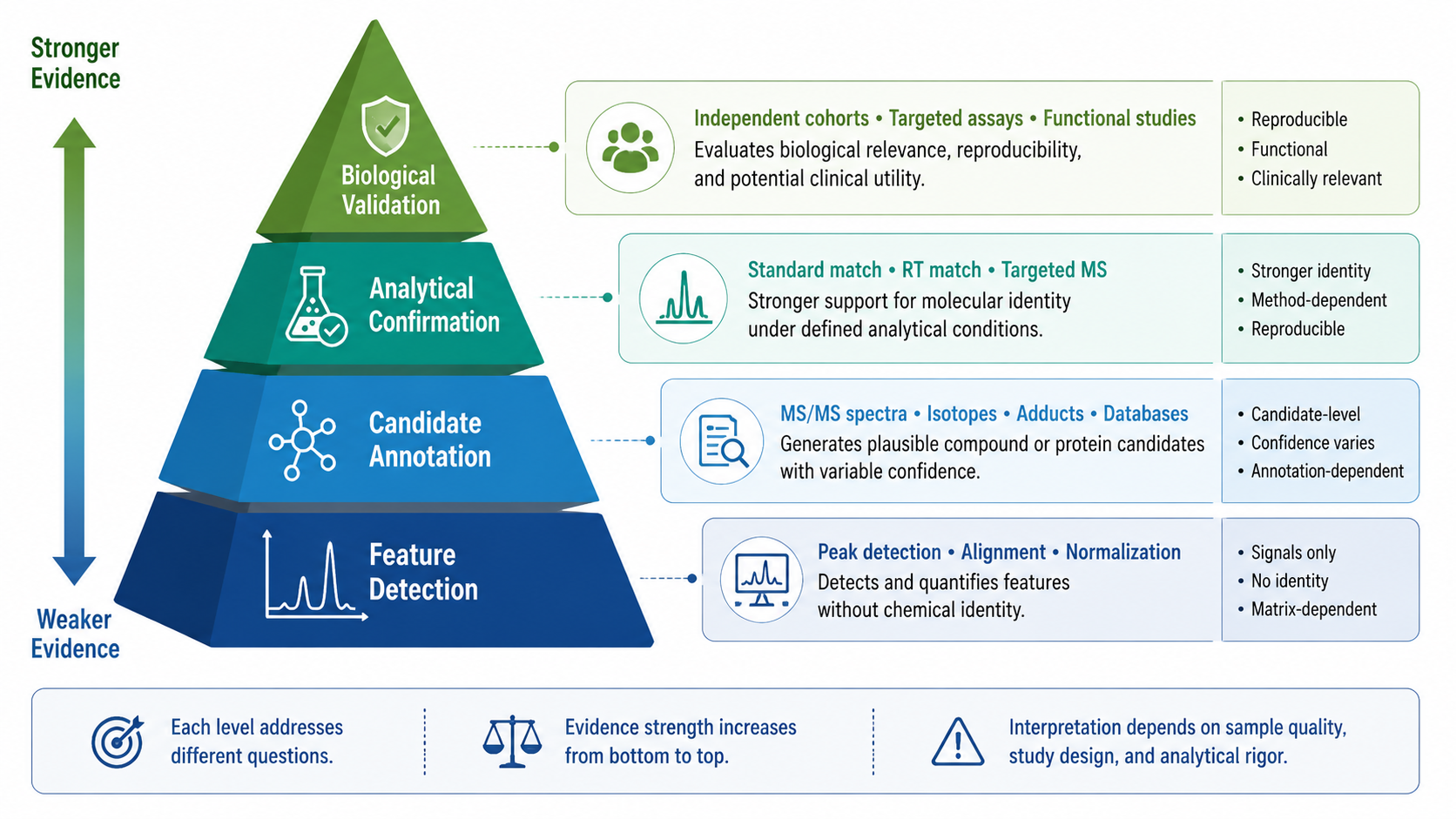
Figure 2. Evidence Hierarchy From EV Proteomics Discovery to Validation
Exosome proteomics analysis is most informative when the starting material, EV preparation status, group design, and expected evidence level are aligned. MtoZ Biolabs supports project evaluation from source samples, pre-isolated EVs, EV protein lysates, or prepared peptides for LC-MS/MS protein identification, quantitative profiling, differential analysis, and project-specific candidate prioritization. Submit your inquiry below for project evaluation.
MtoZ Biolabs, an integrated chromatography and mass spectrometry (MS) services provider.
Related Services
How to order?




