


A Comprehensive Phosphoproteomics Workflow: From Sample Preparation to Data Analysis
Phosphoproteomics represents a crucial subfield of proteomics dedicated to the study of protein phosphorylation and its functional roles within cells. Protein phosphorylation, a common and reversible post-translational modification, involves the addition of phosphate groups to serine, threonine, or tyrosine residues. This process modulates protein activity, subcellular localization, stability, and molecular interactions. In cellular signaling pathways, phosphorylation functions as a molecular switch, governing essential biological processes including cell proliferation, differentiation, apoptosis, and metabolism. Leveraging high-throughput mass spectrometry, phosphoproteomics enables global profiling, quantification, and characterization of phosphorylation site dynamics, thereby uncovering molecular mechanisms underlying disease pathogenesis. This technique holds particular promise in studies of cancer, neurodegenerative disorders, and autoimmune diseases. Furthermore, it plays a pivotal role in drug target identification and biomarker discovery. Through systematic dissection of phosphorylation networks, researchers can construct dynamic signaling pathway maps, facilitating a deeper understanding of cellular responses to external stimuli. Consequently, phosphoproteomics not only advances fundamental life science research but also contributes to clinical diagnostics and precision medicine.
Sample Preparation: Preserving the Integrity of Phosphorylation Modifications
1. Protein Extraction
Phosphorylation sites are highly susceptible to dephosphorylation by endogenous phosphatases. Therefore, phosphatase inhibitors (e.g., NaF, β-glycerophosphate) must be added during sample preprocessing to preserve phosphorylation status. Common sample types, such as cultured cells, tissues, and serum, must be lysed rapidly under cold conditions, typically on ice. Proteins are then extracted using strong chaotropic agents like 8 M urea to ensure efficient solubilization and denaturation.
2. Protein Quantification and Enzymatic Digestion
Total protein concentration must be accurately determined, with the bicinchoninic acid (BCA) assay being the preferred method. Subsequent proteolysis is typically performed using trypsin. Optimizing digestion parameters, such as an enzyme-to-substrate ratio of 1:50 and incubation at 37°C overnight, facilitates the generation of peptides with optimal lengths for mass spectrometric analysis.
Phosphopeptide Enrichment: Enhancing the Detection of Low-Abundance Targets
1. Rationale for Enrichment
Phosphopeptides typically constitute less than 5% of the total peptide population. Without enrichment, these low-abundance species are often masked by high-abundance non-phosphorylated peptides, limiting their detection. Thus, selective enrichment techniques are critical for the success of phosphoproteomic studies.
2. Common Enrichment Strategies
(1) IMAC (Immobilized Metal Affinity Chromatography): Utilizes the affinity of metal ions such as Fe³⁺ or Ga³⁺ for phosphate groups to selectively capture phosphopeptides.
(2) TiO₂-Based Enrichment: Employs metal oxide affinity chromatography to bind phosphate groups, offering high selectivity and reproducibility.
(3) Sequential Enrichment: Combining IMAC and TiO₂ approaches can further enhance phosphopeptide coverage, particularly in complex biological samples.
Mass Spectrometry Detection: Sensitive Identification of Phosphorylation Events
1. LC-MS/MS Analysis
Enriched peptides are first separated using nanoflow liquid chromatography (nanoLC) and then analyzed via high-resolution tandem mass spectrometry, such as the Orbitrap Exploris 480. Fragmentation methods like higher-energy collisional dissociation (HCD) are frequently employed due to their ability to preserve phosphate-specific fragment ions, enabling confident site localization.
2. Data Acquisition Modes
(1) DDA (Data-Dependent Acquisition): Well-suited for building phosphoproteomic atlases and acquiring high-quality spectral data.
(2) DIA (Data-Independent Acquisition): Facilitates large-scale, reproducible, and comprehensive quantification, making it ideal for high-throughput studies.
Bioinformatics Analysis: From Raw Spectra to Biological Insights
1. Data Processing and Quantification
Specialized software tools such as MaxQuant and Proteome Discoverer are widely used for phosphosite localization, signal-to-noise ratio evaluation, and relative quantification. The probability of site localization (e.g., PTM Score) is a critical metric for ensuring data reliability and result confidence.
2. Functional Annotation and Pathway Analysis
(1) GO and KEGG Annotation: Provides insights into biological processes and signaling pathways associated with phosphoproteins.
(2) Motif Analysis: Identifies kinase recognition motifs, aiding in the prediction of upstream regulatory kinases.
(3) Kinase–Substrate Network Reconstruction: Maps kinase–substrate relationships to elucidate signal transduction architecture.
Representative Applications and Scientific Significance
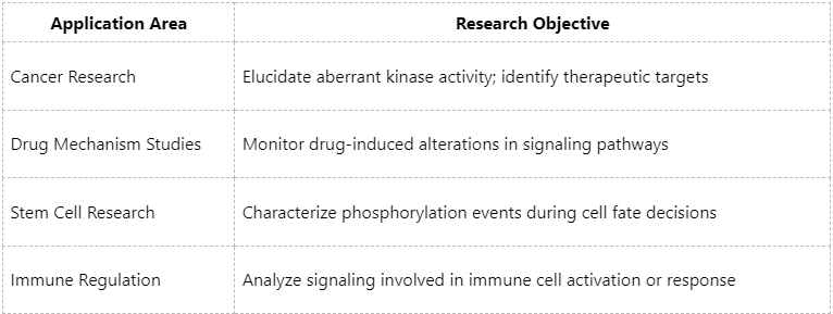
Phosphoproteomics has been extensively applied in cancer biomarker discovery, drug development, and the elucidation of disease mechanisms, establishing itself as a powerful tool at the forefront of precision medicine research.
As a cutting-edge yet technically demanding field, phosphoproteomics offers an indispensable approach for exploring dynamic cellular regulation. Selecting a robust analytical platform and experienced scientific partner is essential for successful project execution. If you are engaged in studies related to signaling pathways, kinase activities, or pharmacological mechanisms, we invite you to collaborate with MtoZ Biolabs. We are committed to delivering high-quality, responsive, and expert services to help you uncover scientific insights from complex phosphoproteomic data.
MtoZ Biolabs, an integrated chromatography and mass spectrometry (MS) services provider.
Related Services
How to order?

