



No. There is no single standard protocol for glutaraldehyde cross-linking. A detailed protocol is available only after the job is named. Electron microscopy or immunohistochemistry wants a fixation path on cells or tissue. A purified-protein oligomer shift wants a much lower dose and a short pulse. A residue-pair search is a different reagent class. Glutaraldehyde is a dialdehyde that polymerizes and builds heterogeneous networks. It is not the usual first choice when the next table must name two linked peptides.
The original six headings remain a usable fixation sequence once that limit is written. Fresh dilution, an amine quench, a PBS wash, a fume hood, and personal protective equipment are kept. Treating 0.1% to 2.5% and minutes-to-hours as the protocol for every protein sample is not. For why this solid is a poor two-protein pair reagent, What Reagents Are Required for In Vitro Cross-Linking of Two Purified Proteins provides a complementary in-vitro reagent page.
Researchers who already know the next table is a residue pair, not a fixed pellet, can also review the MtoZ Biolabs Crosslinking Mass Spectrometry Protein Interaction Analysis Service for project-specific feasibility and analysis planning.
Name the Job Before the Percent
Glutaraldehyde marks primary amines and can keep reacting as the monomer and its polymers add more aldehyde. Commercial stocks are not a single chemical species. A dilution that sat on the bench is not the same reagent as a fresh one. That chemistry is why fixation works and why a defined pair search usually starts elsewhere.
A cell or tissue pellet washed in phosphate-buffered saline (PBS) is the original sample. That start belongs to microscopy and immunohistochemistry. A purified protein already in buffer does not need a media wash as step 1. Copying the cell-wash list onto a two-protein tube is a job error.
If the claim is a lysine-to-lysine pair inside a complex, DSS or BS3 is the usual amine ester. For that site-first choice, How to Select a Crosslinker for XL-MS Structural Studies provides a complementary selection page. For the shared wet order those esters use, What Is the Protocol for Crosslinking-Based Protein Interaction Analysis? provides a complementary protocol page.
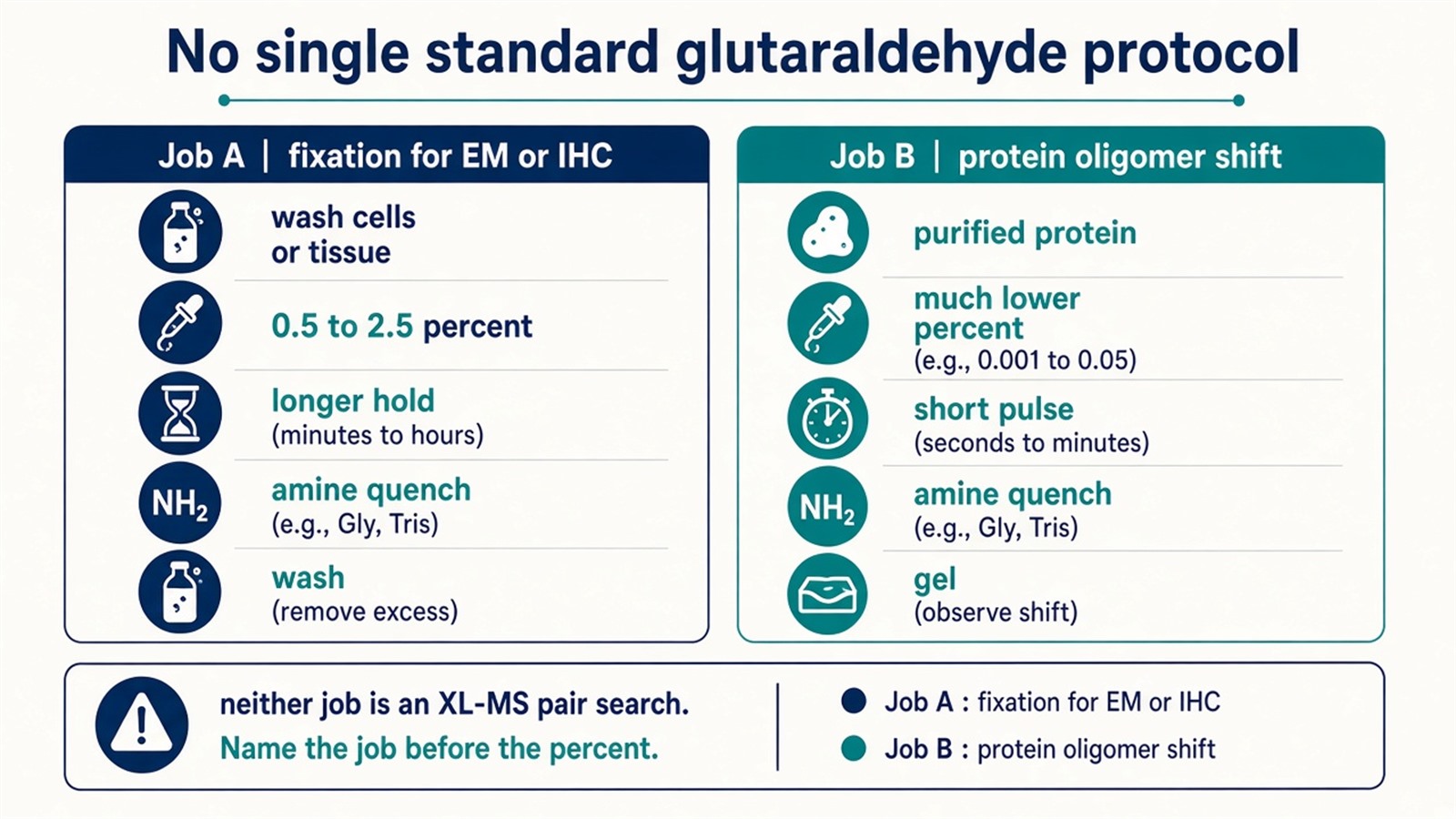
Figure 1. Job A: cells or tissue for EM or IHC. Job B: purified protein for a gel shift. A residue-pair search is not this reagent.
Write the assay before diluting the stock. A published percent range is not a standard protocol.
The Original Six Headings, as Fixation
These headings stay in order when the sample is cells or tissue and the next method is electron microscopy or immunohistochemistry.
1. Sample preparation
Wash to remove residual medium and debris. PBS is a common fixation buffer for aldehydes. It is not a universal protein buffer, and it is not an NHS-ester buffer. A purified-protein oligomer experiment starts from a written protein concentration, not from a flask rinse.
2. Preparation of the glutaraldehyde solution
Dilute a commercial stock immediately before use. The original 0.1% to 2.5% window is a fixation range, not a protein-pair range. Oligomer shifts are often run far below the lower end of that window. Fresh dilution keeps reactivity. It does not make the polymer distribution identical from bottle to bottle.
3. Cross-linking reaction
Cover the sample and hold at room temperature. Minutes can be enough for a thin monolayer. Hours appear in some tissue-fixation recipes. That span is why “typically a few minutes to several hours” cannot be copied onto a purified-protein tube. Over-fixation hardens the pellet and can bury epitopes. Under-fixation leaves the sample unstable in later solvent.
4. Quenching
Add an amine such as Tris or glycine to consume leftover aldehyde. The quench stops further marking. It does not reverse networks that already formed. The original “15 minutes to several hours” belongs next to the pulse, not as a second copy of the reaction time after the stop.
5. Post-treatment
Wash with PBS or the same buffer to remove residual glutaraldehyde. Several washes reduce leftover aldehyde. They do not purify a protein complex.
6. Subsequent analysis
Electron microscopy and immunohistochemistry remain the original readouts for this path. They ask whether morphology or an epitope survived the fixation. They do not report residue pairs. A stained gel after a low-dose protein pulse is the oligomer-shift readout. It is a mass shift, not a site.
A no-glutaraldehyde arm is processed through the same quench and wash. Changing only the treated sample mixes a handling effect with a reagent effect.
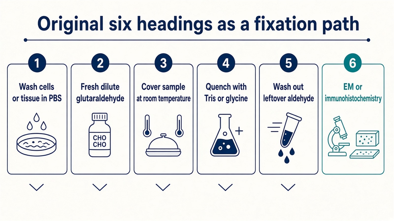
Figure 2. Wash, fresh dilute, cover, amine quench, wash, then EM or IHC. That is fixation. It is not a standard protein-pair protocol.
What a Detailed Protocol Must Still Name
A usable write-up names the job, the sample state, the percent or millimolar dose, the buffer, the time, the quench, and the assay. Without those fields the six headings are a checklist, not a detailed protocol. Hazard controls stay on the same card. Glutaraldehyde is handled in a ventilated fume hood with eye, skin, and respiratory protection. That note in the original page remains.
Stabilizing proteins and other macromolecules is the intent of a covalent network. The protocol does not grant a native-state architecture or a complete interface. Intra-molecular and inter-molecular links both form, and polymers add extra reach that is not a defined spacer.
If the next sentence is still a residue-pair table, switching the solid to DSS or BS3 after a failed glutaraldehyde gel does not convert these six headings into an XL-MS method. For the three readouts that use a defined amine ester, What Are the Specific Experimental Methods for Cross-Linking Protein Interaction Analysis provides a complementary methods page.
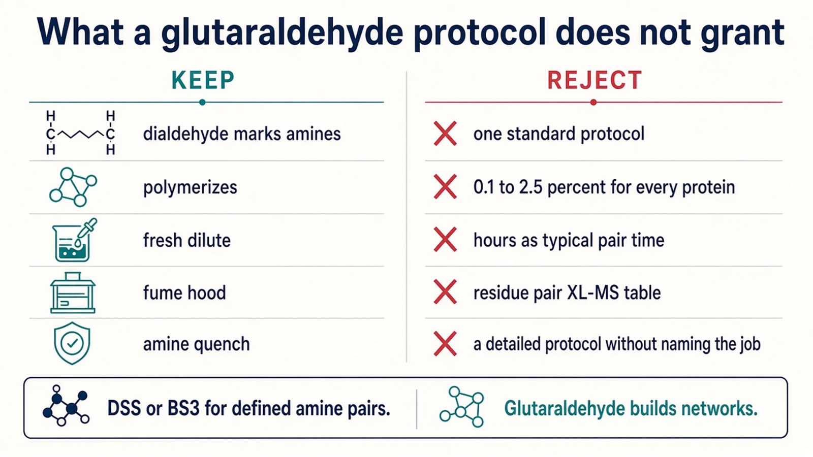
Figure 3. Keep: dialdehyde, polymerization, fume hood, Tris or glycine stop. Not granted: one standard percent, hours as a pair time, or a residue-pair table.
Experimental Limits
A glutaraldehyde protocol reduces a sample to one job, one dose, one quench, and one assay. Diagnostic completeness of every macromolecular contact sits outside ordinary research reporting. A detailed protocol that never names the job is still not available.
Project Planning and Service Scope
A usable plan names the job, the percent, the quench, and the claim that the network is allowed to support, before the first dilution. MtoZ Biolabs scopes crosslinking MS with Orbitrap-class LC-MS/MS. Interpretability still follows those arms, not a standard-glutaraldehyde slogan.
Once the next sentence is a cellular partner list rather than a fixed pellet or a residue pair, the project should not be forced through this six-step list. Researchers who need that pellet can review the MtoZ Biolabs Protein-Protein Interaction Analysis Service for project-specific sample evaluation, feasibility assessment, and workflow planning.
Frequently Asked Questions
1. Is there a standard protocol for glutaraldehyde cross-linking?
No. Fixation and a protein oligomer shift do not share one recipe.
2. Is a detailed protocol available?
Yes, after the job, the dose, the time, the quench, and the assay are named.
3. Is 0.1% to 2.5% the usual protein-crosslinking range?
That window is a fixation range. Oligomer shifts often use a much lower dose.
4. Should leftover glutaraldehyde be quenched with Tris?
An amine such as Tris or glycine consumes leftover aldehyde. Hours after the pulse are not the quench.
5. Is glutaraldehyde the standard reagent for XL-MS pair search?
No. It builds heterogeneous networks. Defined amine pairs usually start with DSS or BS3.
Reference
- Migneault I., Dartiguenave C., Bertrand M.J., Waldron K.C. (2004). Glutaraldehyde: behavior in aqueous solution, reaction with proteins, and application to enzyme crosslinking. BioTechniques, 37, 790-802. https://doi.org/10.2144/04375RV01
- Klockenbusch C., Kast J. (2010). Optimization of formaldehyde cross-linking for protein interaction analysis of non-tagged integrin beta1. J. Biomed. Biotechnol., 2010, 927585. https://doi.org/10.1155/2010/927585
- Leitner A., Walzthoeni T., Kahraman A., et al. (2014). Probing native protein structures by chemical cross-linking, mass spectrometry, and bioinformatics. Nat. Protoc., 9, 120-135. https://doi.org/10.1038/nprot.2013.168
- O'Reilly F.J., Rappsilber J. (2018). Cross-linking mass spectrometry: methods and applications in structural, molecular and systems biology. Nat. Struct. Mol. Biol., 25, 1000-1008. https://doi.org/10.1038/s41594-018-0147-0
- Yu C., Huang L. (2018). Cross-linking mass spectrometry: an emerging technology for interactomics and structural biology. Anal. Chem., 90, 144-165. https://doi.org/10.1021/acs.analchem.7b04431
