



Protein-protein interaction analysis asks which proteins remain with a bait after capture and matched controls. Single-cell CUT&Tag does not answer that question. It asks which genomic regions are occupied by a chromatin protein or histone mark in individual cells after antibody-guided tagmentation, negative-control contrast, and peak or cell-level ranking. The path fits nuclei or cells when a CUT&Tag-competent antibody exists and the claim is occupancy, not protein-protein contact or functional regulation (Kaya-Okur et al. 2019). Prefer this path when cell-type heterogeneity is the written question. Reassess when the antibody is Western-only, IgG or no-primary is missing, or peaks are written as PPI edges.
Researchers who already have a chromatin target, antibody status, and a written occupancy claim can also review the MtoZ Biolabs CUT&Tag Analysis Service for project-specific single-cell feasibility.
Which Experimental System Can Support Single-Cell CUT&Tag
Single-cell CUT&Tag is a protein-chromatin occupancy assay. An antibody binds a histone mark or a chromatin-associated protein in permeabilized cells or nuclei. A protein A/G-Tn5 fusion then tagments nearby DNA. Sequencing ranks those fragments by cell. Bulk CUT&Tag chemistry is the same capture class. Single-cell isolation is the added system constraint, not a more complete interaction method.
The system is readable when three things are named. First, the target is a chromatin feature, not a soluble PPI bait. Second, the antibody is plausible under CUT&Tag conditions. Western performance does not establish that. Third, nuclei or cells can be isolated without destroying the epitope or flooding the library with free DNA. Droplet, FACS, or plate isolation can all finish the path when those three conditions hold. None of them converts occupancy into protein-protein interaction analysis.
How bulk CUT&Tag is designed is covered in Beginner’s Guide: How to Start CUT&Tag Analysis. When the written sentence is which proteins remain with a protein bait, use Protein-Protein Interaction Analysis instead of single-cell CUT&Tag.
The system is a poor first choice when no CUT&Tag-competent antibody exists, when the material cannot yield intact nuclei, or when the claim is reconstituted protein-protein binding or a complete regulatory network.
Single-cell CUT&Tag reports candidate chromatin occupancy in named cells. It does not certify a protein-protein contact.
Negative Controls Before Any Peak Is Kept
Negative controls are the denominators of single-cell CUT&Tag. They are not optional after one cluster looks clean.
IgG or no-primary arms show antibody-independent tagmentation. They must see the same permeabilization, Tn5 activation, and library recipe as the target antibody. A longer wash on the target only, or a different Tn5 window on the control, makes enrichment unreadable. Beads-only from an IP-MS notebook does not replace IgG here.
A no-Tn5 or killed-enzyme arm, when used, shows carryover DNA that was not created by targeted tagmentation. Spike-in or barcode-balanced controls can compare batches. They do not replace IgG. Input DNA from ChIP-seq is not automatically a single-cell CUT&Tag control. If an input-like arm is kept, it must be processed through the same single-cell barcode path.
Cell-hashing or unused barcodes can flag empty droplets and collisions. They do not prove that a peak in a real cell is target-specific.
System check |
What a pass looks like |
What the analysis cannot say |
|---|---|---|
Experimental system |
Named chromatin target, CUT&Tag-competent antibody, intact nuclei or cells |
A PPI bait recovered from a lysate |
Negative control |
IgG or no-primary on the same Tn5 recipe |
Every remaining peak is target-specific |
Candidate screening |
Occupancy ranked versus those controls in named cells |
An unfiltered fragment table as a network |
Result boundary |
Candidate occupied regions under those arms |
Protein-protein contact or gene regulation |
A practical path is:
- Write the occupancy sentence the cell-by-peak table must support.
- Confirm the antibody recovers the intended chromatin target in a pilot.
- Process IgG or no-primary with the same tagmentation recipe.
- Rank only regions or cells that pass that contrast.
- Keep a shortlist for an orthogonal RNA, motif, or perturbation assay.
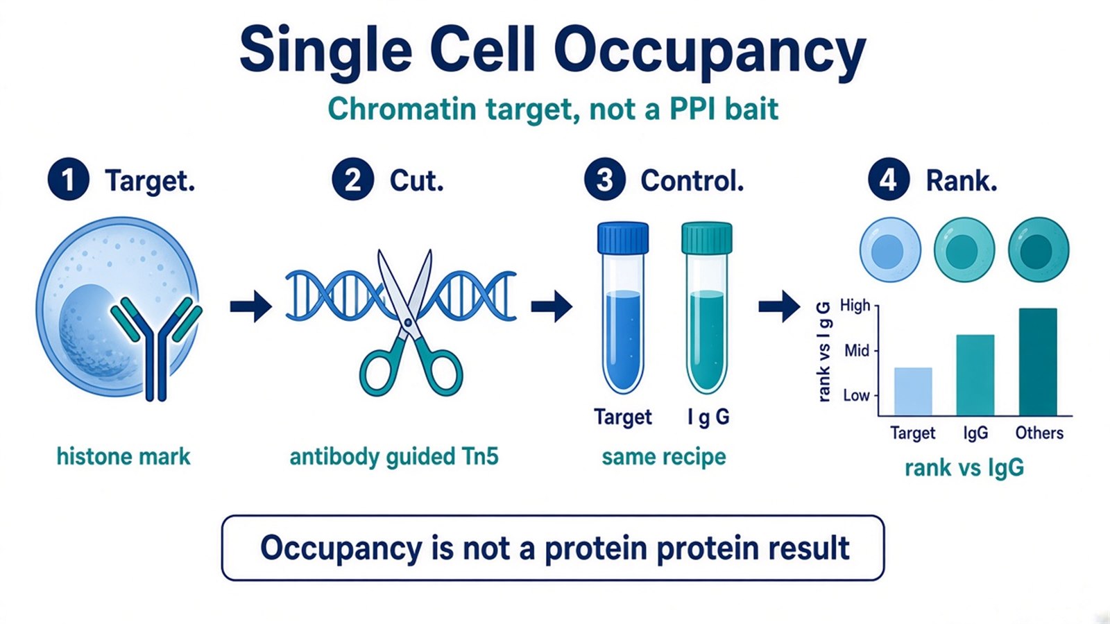
Figure 1. Single-cell CUT&Tag starts after the chromatin target is named. A droplet cartoon is not an interaction result.
Screening Candidate Occupied Regions
Candidate screening starts after the target antibody produces fragments above the matched control. If IgG or no-primary libraries look like the target library, stop ranking cell types.
A peak or a cell-level enrichment score is a contrast against the control recipe. It is not a partner protein. Sparse single-cell counts can hide a real mark or inflate a noisy barcode. Depth, cell number, and batch correction can change detection. They do not turn occupancy into PPI, and they do not assign a gene as a regulated target.
Do not write a cluster-specific peak set as a protein interaction network. Do not write a histone-mark domain as proof that a transcription factor sits at every bin. Do not write nearest-gene annotation as regulation.
How occupancy ranks are turned into a peak table is covered in How to Perform CUT&Tag Peak Calling and Functional Annotation?.
Candidate screening ranks occupied regions versus matched negative controls. It does not certify a binary protein-protein or protein-DNA contact.
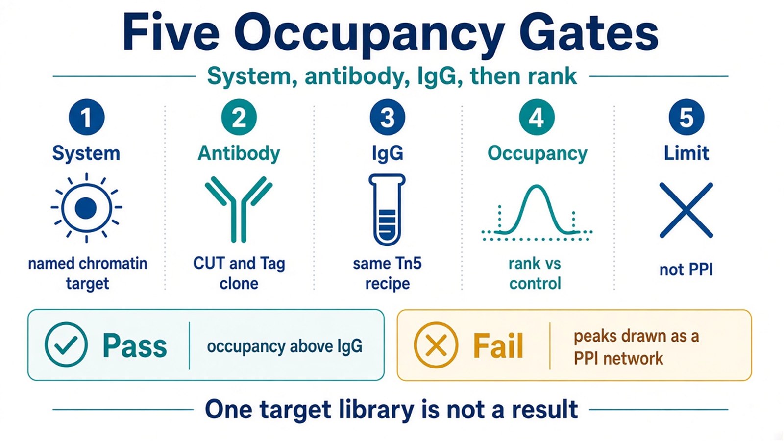
Figure 2. Screening is a control outcome. Extra sequencing does not replace IgG or no-primary.
Result Boundaries and Common Biases
The common bias is writing single-cell CUT&Tag as high-sensitivity protein interaction analysis because an antibody and a sequencing table were used. Another bias is skipping IgG because material is limited, then drawing leftover fragments as cell-type-specific regulation. Over-tagmentation can fill every barcode with background. Under-permeabilization can hide the epitope.
Do not write a successful target library as proof that remaining peaks contact the protein. Do not write a negative cell as proof that the mark is absent in that lineage. Absence can mean the wrong antibody, lost nuclei, barcode collision, or a mark below the sparse detection floor. Do not write a CUT&Tag peak as a functional regulatory site.
Prefer single-cell CUT&Tag when occupancy must be split by cell state. Prefer bulk CUT&Tag when the question is a population-average map and material is not limiting. Prefer protein-protein interaction analysis when the next sentence is which proteins remain with a protein bait.
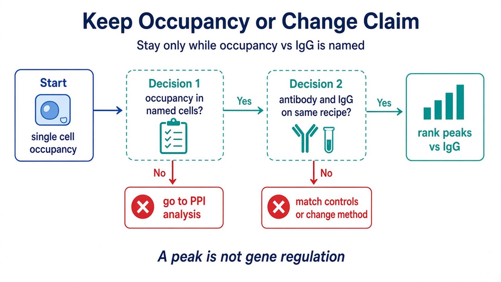
Figure 3. Keep single-cell CUT&Tag only while occupancy versus IgG can be named. A cluster map is not a completed interaction analysis.
Once the target, antibody status, isolation class, negative controls, and the planned ranking are defined, the project can be scoped as single-cell CUT&Tag occupancy analysis. Researchers can review the MtoZ Biolabs CUT&Tag Analysis Service for sample evaluation and path planning.
Frequently Asked Questions
1. Does single-cell CUT&Tag analyze protein-protein interactions?
No. It maps chromatin occupancy in named cells. PPI analysis recovers proteins that remain with a protein bait.
2. Can I skip IgG because single-cell material is limited?
No. IgG or no-primary must see the same Tn5 recipe. One target library is not a CUT&Tag result.
3. Does a peak mean the protein regulates that gene?
No. A peak is occupancy versus control. Regulation needs a perturbation or other orthogonal assay.
4. Is bulk CUT&Tag the same as single-cell CUT&Tag?
No. The chemistry is the same occupancy class. Single-cell isolation changes sparsity and control design, not the written claim.
5. When should this analysis move to a PPI or pair assay?
Move when the next written sentence is protein-protein contact, direct DNA contact, or affinity. Keep the occupancy shortlist as the input, not as the proof.
Reference
- H.S. Kaya-Okur, S.J. Wu, C.A. Codomo, E.S. Pledger, T.D. Bryson, J.G. Henikoff, K. Ahmad, S. Henikoff (2019). CUT&Tag for efficient epigenomic profiling of small samples and single cells. Nat. Commun., 10, 1930. https://doi.org/10.1038/s41467-019-09982-5
