



Protein–protein interactions (PPIs) are the associations through which proteins carry out signaling, metabolic regulation, cell-cycle control, and many disease-associated processes. A single-protein abundance value rarely explains function. PPI analysis is used to place a protein in a pathway, to map how a network is disrupted in disease, and to ask whether an interface can be modulated by a compound or a degrader. Those three biological jobs do not share one analytical technique. Co-immunoprecipitation (Co-IP), yeast two-hybrid (Y2H), affinity purification–mass spectrometry (AP-MS), quantitative interactomics, and crosslinking mass spectrometry (XL-MS) answer different parts of the same field.
In the cellular microenvironment, proteins rarely act in isolation. Systematic interrogation of PPI networks is therefore a central route in functional proteomics, biomarker follow-up, and mechanistic studies of drug action. Biological significance is a claim about what the interaction does. The technique must still match the physical event being measured: a wash-stable complex, a binary pair in yeast, a condition-dependent occupancy change, or a contact site.
Researchers who already have a defined protein, pathway, or project objective can also review the MtoZ Biolabs Protein-Protein Interaction Analysis Service for project-specific feasibility and analysis planning.
Biological Significance of Protein–Protein Interactions
1. Protein function and signaling mechanisms
Expression of one protein is often insufficient to assign its role. PPI-focused experiments can show which signaling pathway a protein enters, which upstream or downstream partners it uses, and whether it acts with or against other components. The apoptotic functions of p53, for example, are interpreted through contacts with transcription factors and ubiquitin ligases, not through p53 abundance alone. That example illustrates pathway context. It does not make every recovered partner a functional regulator.
2. Disease-mechanism analysis and target nomination
Many cancers and neurodegenerative diseases involve disrupted PPI networks rather than a single enzyme active site. Aberrant contacts between Tau and microtubule-associated proteins are one widely discussed step in Alzheimer-related models. Profiling PPI perturbations can nominate therapeutic targets and flag pathways that may be affected if an interface is later blocked. Those lists remain experimental hypotheses. They are not diagnoses and they do not, by themselves, predict clinical adverse events.
3. Drug-development strategies at PPI interfaces
Modulating a PPI interface is a distinct drug-discovery direction from classical active-site inhibition. Agents that disrupt, reshape, or reassemble a complex, including proteolysis-targeting chimeras (PROTACs) as one strategy class, act on proximity or occupancy rather than on catalytic turnover. Interface drugs still require a defined complex, a measurable occupancy change, and later orthogonal pharmacology. A PPI table is not a medicinal-chemistry starting point until the edge is validated.
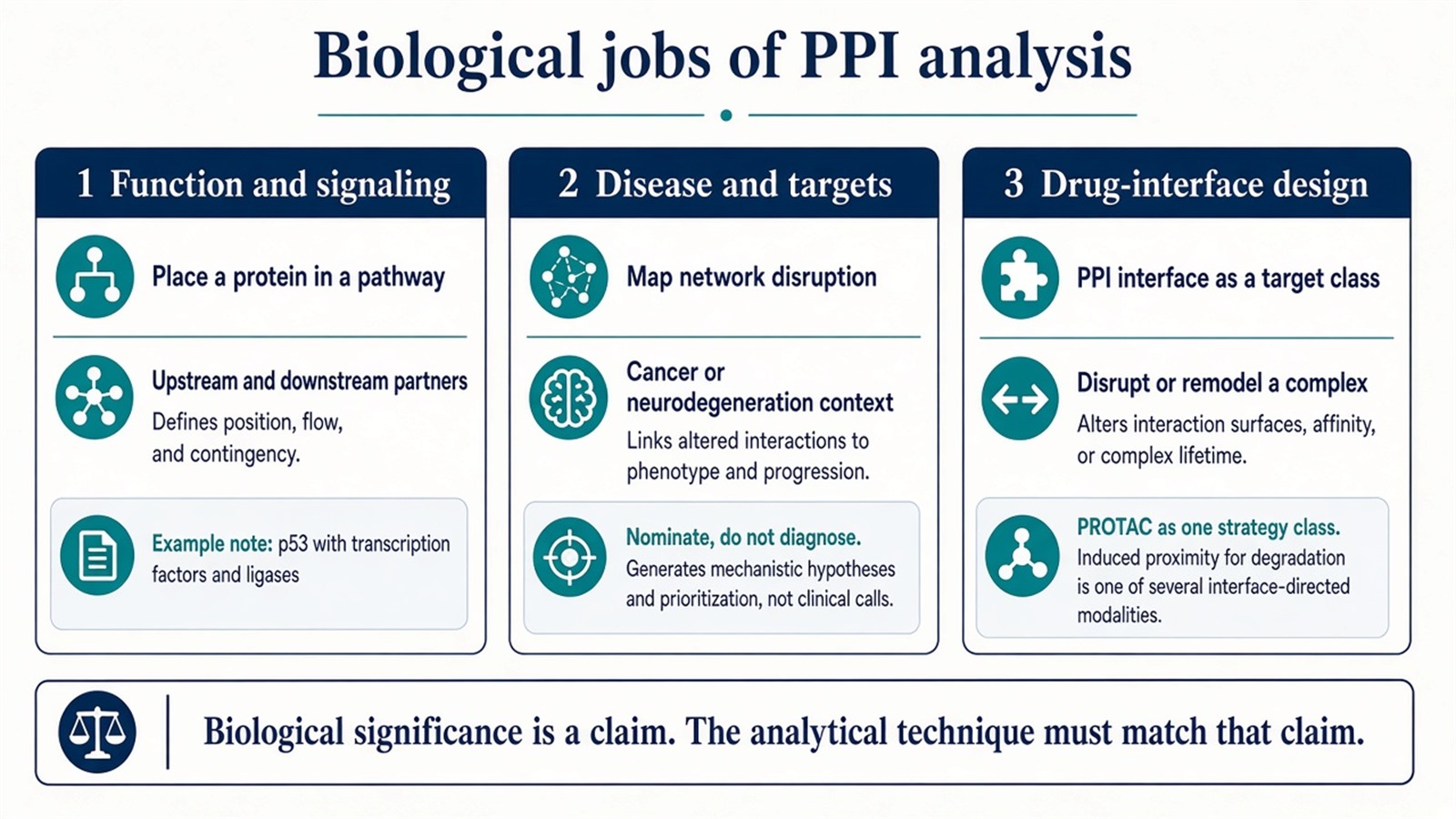
Figure 1. PPI analysis supports function assignment, disease-network nomination, and interface-directed drug design; each job still needs a matching analytical technique.
Biological significance names the next sentence. It does not rank Co-IP, Y2H, or AP-MS as universally more important.
Analytical Techniques for PPI Studies
1. Co-immunoprecipitation (Co-IP)
Co-IP uses an antibody to enrich a target protein and any partners that survive lysis and washing. The eluate is read by Western blot or by mass spectrometry. It is a common validation method for stable complexes, not a gold standard for every PPI claim. The assay is biased toward abundant, wash-stable contacts, depends on antibody specificity, and is low throughput for omics-scale screens.
2. Yeast two-hybrid (Y2H)
Y2H is a transcriptional-activation screen for binary pairs and remains useful for first-pass network construction. False-positive rates can be high. Membrane proteins and interactions that need a multi-subunit assembly or mammalian modifications are often out of scope.
3. Affinity purification–mass spectrometry (AP-MS)
AP-MS enriches a tagged or antibody-captured complex and identifies co-purified proteins by high-resolution LC-MS/MS under near-physiological lysis. It preserves a cellular context that Y2H cannot and supports higher partner coverage than a single Western. Specificity still depends on matched empty-tag or bead-only controls. Tagging and overexpression can create non-physiological partners.
MtoZ Biolabs runs AP-MS on an Orbitrap Exploris 480-class platform with a low-background enrichment workflow. Low-abundance recovery still depends on bait expression, wash stringency, and control filtering, not on the platform name alone.
4. Quantitative interactomics
SILAC combined with AP-MS compares the same bait-associated proteins across treatments and can show how an interaction landscape shifts. Label-free interactomics is used when clinical or other samples cannot be metabolically labeled; it compares MS signal under a matched capture chemistry. Both report relative occupancy. Neither reports a Kd.
5. Crosslinking mass spectrometry (XL-MS)
Chemical crosslinkers stabilize spatial architecture so that mass spectrometry can assign binding-site and conformational constraints. XL-MS is used for dynamic or transient contacts that washing would lose, for structural modeling support, and for conformational change around an interface. Coverage is sparse relative to a complete surface.
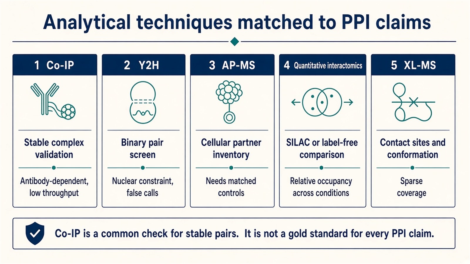
Figure 2. Co-IP validates stable pairs, Y2H screens binary contacts, AP-MS inventories cellular partners, quantitative interactomics compares occupancy, and XL-MS maps sites.
For claim-first method selection across these families, How to Analyze Protein–Protein Interactions provides a more detailed explanation of when Co-IP, AP-MS, Y2H, and biophysical assays fit.
Biological question |
Technique that often fits |
Reassess |
|---|---|---|
Is this known pair present as a wash-stable complex? |
Co-IP, optionally with MS |
No usable antibody, or the contact is transient |
Which binary partners can reconstitute in a screen? |
Y2H |
Membrane or PTM-dependent contacts |
Which proteins co-purify with this bait in cells? |
AP-MS with matched controls |
Only one prey needs a Western |
How does occupancy change after treatment? |
SILAC or label-free interactomics |
A Kd on purified proteins is required |
Where do the chains contact? |
XL-MS |
Only a partner list is needed |
Can this interface be a drug-design starting point? |
Validated edge plus later pharmacology |
An unscored AP-MS table alone |
From Single-Pair Validation to Functional Proteomics
PPI work is moving from one-edge confirmation toward systematic, condition-aware maps. Transcriptomics, epigenomics, and metabolomics can add regulatory context around a scored interactome. They do not replace experimental PPI evidence. Omics-scale modeling is still limited by the capture chemistry: a Y2H map, an AP-MS occupancy table, and an XL-MS site set are not the same network.
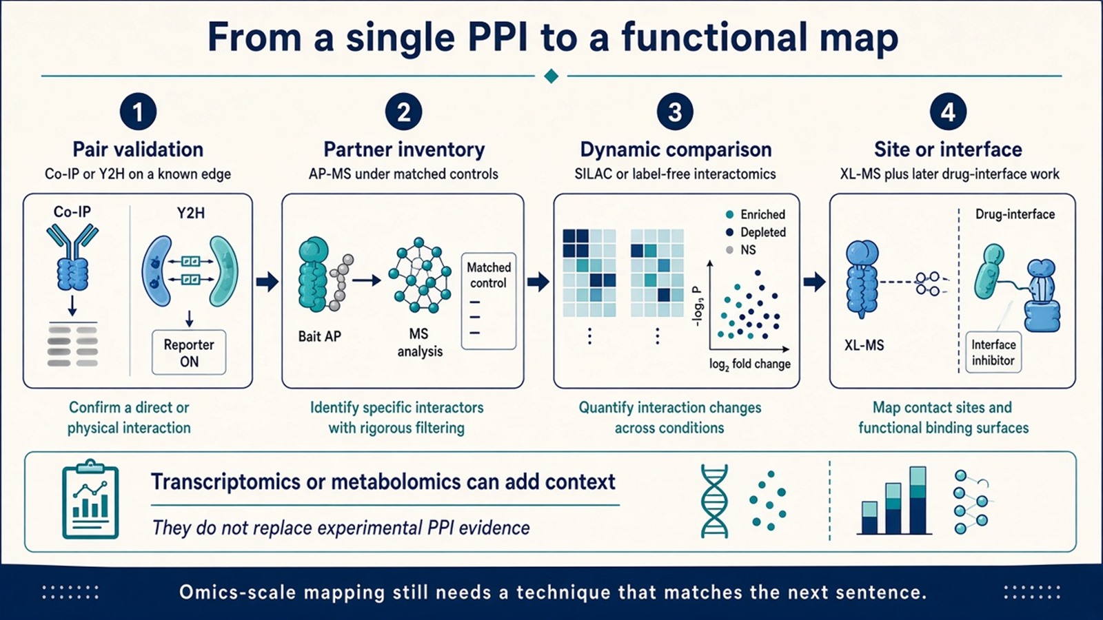
Figure 3. Functional PPI maps are built by sequencing validation, partner inventory, occupancy comparison, and site-level work; multi-omics context does not replace those measurements.
For MS-centered identification and quantification design, How to Identify Protein–Protein Interactions Using Mass Spectrometry provides a more detailed explanation of AP-MS, XL-MS, and proximity-labeling evidence.
Experimental Limits
Co-IP is not a universal gold standard. Y2H and AP-MS will disagree when they measure different events. SILAC and label-free tables remain relative. XL-MS sites constrain geometry; they do not finish a structure. Disease examples such as p53 or Tau illustrate pathway logic. They do not validate a new bait. Diagnostic, companion-diagnostic, or clinical-translation claims sit outside ordinary research PPI reporting unless a separately validated assay framework is in place.
Project Planning and Service Scope
A usable PPI plan names the biological job, the physical event, and the technique. MtoZ Biolabs can scope AP-MS partner identification, SILAC or label-free differential interactomes, XL-MS conformational characterization, and pathway-level interpretation after the experimental table is closed. Publishable use still depends on controls and a declared score, not on a quality-control slogan.
Once the biological claim and technique are defined, the project can be scoped as a PPI study. Researchers can review the MtoZ Biolabs Protein-Protein Interaction Analysis Service for project-specific sample evaluation, feasibility assessment, and workflow planning.
Frequently Asked Questions
1. Is Co-IP the gold standard for all protein–protein interactions?
No. Co-IP is a common check for wash-stable complexes when a usable antibody exists. Transient, membrane, or omics-scale questions need other methods.
2. Can PPI analysis explain protein function without abundance data?
It can place a protein among partners. Function still needs perturbation or orthogonal assays. Abundance and interaction are separate measurements.
3. Do AP-MS results justify a PPI-interface drug program?
Not by themselves. Interface design needs a validated edge, a measurable occupancy or structural constraint, and later pharmacology.
4. When is quantitative interactomics required instead of a single AP-MS list?
When the biological sentence is about change after treatment, genotype, or tissue state, not only about which proteins co-purify once.
5. Does multi-omics integration prove a PPI network?
No. Transcriptomic or metabolomic layers add context. Sample-level PPI evidence remains the Co-IP, Y2H, AP-MS, or XL-MS experiment.
Reference
- Berggard T., Linse S., James P. (2007). Methods for the detection and analysis of protein-protein interactions. Proteomics, 7, 2833-2842. https://doi.org/10.1002/pmic.200700131
- Gingras A.C., Gstaiger M., Raught B., Aebersold R. (2007). Analysis of protein complexes using mass spectrometry. Nat. Rev. Mol. Cell Biol., 8, 645-654. https://doi.org/10.1038/nrm2208
- Fields S., Song O. (1989). A novel genetic system to detect protein-protein interactions. Nature, 340, 245-246. https://doi.org/10.1038/340245a0
- Roux K.J., Kim D.I., Raida M., Burke B. (2012). A promiscuous biotin ligase fusion protein identifies proximal and interacting proteins in mammalian cells. J. Cell Biol., 196, 801-810. https://doi.org/10.1083/jcb.201112098
- Bekes M., Langley D.R., Crews C.M. (2022). PROTAC targeted protein degraders: the past is prologue. Nat. Rev. Drug Discov., 21, 181-200. https://doi.org/10.1038/s41573-021-00371-6
