



To detect a protein in cell experiments and determine whether it is expressed, the first decision is what “expressed” must mean in that experiment: presence of a named protein, relative amount, subcellular localization, or an open protein inventory. Western blot, immunofluorescence, and ELISA report a named protein when a suitable antibody exists. Mass spectrometry identifies proteins from digested cell extracts without requiring an antibody for each target, and it can quantify them only under a declared design. RT-qPCR measures mRNA and is not a protein assay. These techniques are therefore not interchangeable answers to the same question.
A band, a fluorescent image, an optical-density value, a transcript fold-change, and a peptide-spectrum match support different claims. Mixing those claims is the usual reason a detection experiment is later hard to interpret.
Researchers who already have defined samples, comparison groups, or a specific project objective can also review the MtoZ Biolabs Protein Identification Service for project-specific feasibility and analysis planning.
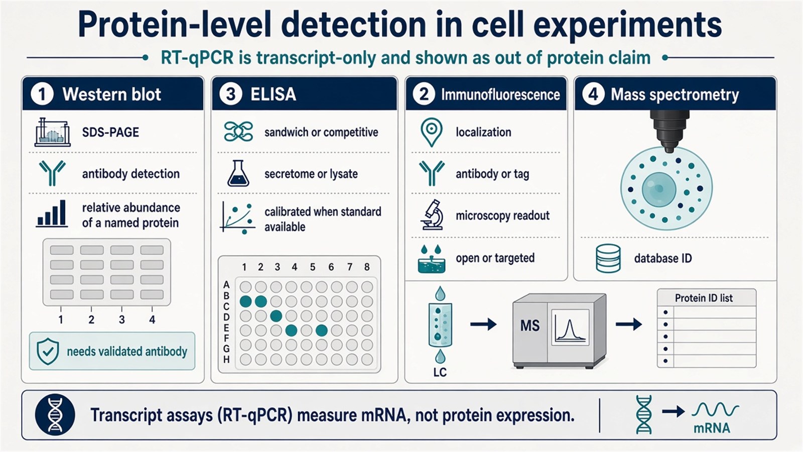
Figure 1. Protein-level detection in cell experiments commonly uses Western blot, immunofluorescence, ELISA, or mass spectrometry; RT-qPCR is a transcript assay and does not prove protein abundance.
Detecting protein expression means measuring the protein under a stated claim, not inferring it from any convenient signal.
How to Choose a Detection Method
Choose the method from the scientific claim, the antibody status, and whether the protein list is closed. Antibody methods are efficient when the target is named and the reagent is adequate. Mass spectrometry is the route when the question is identity across many proteins, when no antibody exists, or when a gel band or immunoprecipitate still needs sequence evidence. RT-qPCR belongs in the experiment when the question is transcription, not protein presence.
|
Experimental need |
Often use |
Reassess |
|---|---|---|
|
Named protein presence and apparent molecular weight |
Western blot |
Treating any band as identity without size and control lanes |
|
Named protein amount across many lysates |
ELISA |
Using ELISA to prove molecular weight or localization |
|
Localization inside intact cells |
Immunofluorescence |
Reading fluorescence intensity as calibrated protein amount |
|
Open protein list, no antibody, or sequence evidence |
Mass spectrometry |
Using MS as a drop-in replacement for one Western blot |
|
Transcript abundance of the target gene |
RT-qPCR |
Inferring protein expression from mRNA alone |
Prefer Western blot when the claim needs a size-resolved antibody signal. Prefer ELISA when the claim is amount of a named antigen under a standard curve. Prefer immunofluorescence when the claim is where the protein sits. Prefer mass spectrometry when the claim is identification or a proteome-level comparison. Prefer RT-qPCR only for mRNA.
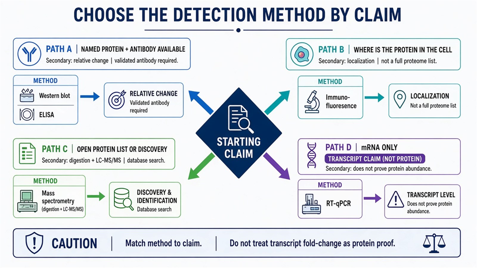
Figure 2. Method choice follows the claim: antibody methods for a named protein, immunofluorescence for localization, mass spectrometry for an open protein list, and RT-qPCR for mRNA only.
Western Blot
Western blot is a common method to detect proteins in cell lysates. Cell proteins are first separated by gel electrophoresis, then transferred to a membrane. A specific antibody is used to detect the target protein. If the target protein is present at a detectable level and the antibody binds, a band appears at the corresponding position on the membrane.
That band supports presence of an immunoreactive species at an apparent molecular weight. It does not, by itself, prove that the protein is correctly folded, active, or the only species recognized by the antibody. Loading controls, transfer quality, and antibody validation determine whether the result can be compared across samples. Relative densitometry is a comparison under matched load, not an absolute concentration.
Western blot remains the usual first protein-level check for knockdown, overexpression, and treatment studies when an antibody exists. It is a poor proteome survey and a poor localization assay.
For a closer comparison of Western blotting and ELISA as protein quantification choices, researchers can refer to Principles and Steps of Western Blotting, Differences with ELISA, and Choosing the Right Method for Protein Quantification for more detailed guidance.
Immunofluorescence
Immunofluorescence uses specific antibodies and fluorescently labeled secondary antibodies to observe protein expression and localization in cells. Cells are fixed on a slide and stained with a primary antibody targeting the protein. The secondary antibody is usually fluorescently labeled. Under a fluorescence microscope, the distribution of the labeled protein within the cells can be observed.
The method answers where an immunoreactive signal sits after fixation and permeabilization. It does not automatically report bulk expression in a lysate, and fixation can hide or create apparent localization. Controls for secondary-only staining, isotype background, and, where possible, genetic knockout or knockdown are part of the claim. Quantitative imaging can rank relative intensity under one protocol. It is not a calibrated protein concentration unless that calibration is separately designed.
Use immunofluorescence when localization is the experimental question. Use a lysate assay when the question is amount in an extract.
RT-qPCR
RT-qPCR measures mRNA expression. It can serve as an indirect indicator of whether a gene is transcribed, but it does not detect the protein. By quantifying mRNA levels of the target gene, researchers obtain transcript abundance, not protein copies. The practical steps are extraction of total RNA from cells, reverse transcription into cDNA, and real-time quantitative PCR.
mRNA and protein abundance often diverge because of translation control, protein turnover, and trafficking (Vogel and Marcotte 2012). A transcript increase can occur without a matching protein increase, and a protein can persist after mRNA has fallen. RT-qPCR is therefore a companion transcription assay, not a substitute Western blot, ELISA, or mass spectrometry result.
ELISA
ELISA uses enzyme-linked detection and specific antibodies to quantify target proteins. Protein is extracted from cells and adsorbed onto an enzyme-linked plate, or captured in a sandwich format. Specific antibodies and enzyme-conjugated secondary reagents are then used for detection. The protein level is quantified through an enzyme-substrate reaction and optical density measurements, usually against a standard curve.
ELISA fits named-protein amount when the antibody pair, matrix, and curve are adequate. It does not report molecular weight, and it does not identify unexpected proteins in the extract. Hook effects, incomplete extraction, and off-target antibody binding remain experimental limits. For a closed antigen and many samples, ELISA is often more practical than densitometry of Western blot bands. For identity or size, it is not.
Mass Spectrometry
Mass spectrometry identifies and, under a separate design, quantifies proteins in cells without requiring a pre-chosen antibody for each target. After cell extracts are digested with a protease to generate peptides, a mass spectrometer detects those peptides. The resulting spectra are matched to a protein database, which helps determine whether the target protein, and often many other proteins, is present in the cells.
Bottom-up LC-MS/MS is the usual route for complex cell lysates: peptides are separated by liquid chromatography, fragmented, and assigned by database search (Aebersold and Mann 2016). Identification supports presence under the stated false-discovery and uniqueness filters. Quantification is a different claim. Label-free or isobaric workflows can compare the same protein across cell states. Absolute amount still needs matched standards. A single peptide-spectrum match is weak evidence; protein inference from more than one unique peptide is the more defensible reporting unit.
Mass spectrometry is the method of choice when no antibody exists, when a gel band or immunoprecipitate needs sequence identity, or when the experiment must move from one protein to a cellular protein matrix. It is not a faster Western blot for a single well-validated antibody target, and it does not by itself provide immunofluorescence-style localization.
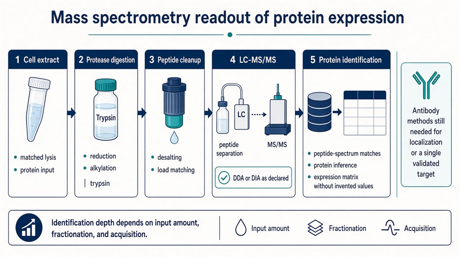
Figure 3. Mass spectrometry detection of protein expression typically proceeds from a cell extract through protease digestion, cleanup, and LC-MS/MS to database-supported protein identification.
A peptide-spectrum match supports protein presence only under the digestion, search, and filter rules used for that experiment.
For cell-wide identification and quantitative comparison after a detection question has expanded beyond one antibody target, researchers can refer to the MtoZ Biolabs Cellular Proteomics Service for more detailed project scoping.
Experimental Limits and Design Boundaries
Antibody methods inherit antibody specificity, epitope access, and linear range. A Western blot band at the wrong size, an ELISA signal without a parallel size check, or immunofluorescence without genetic or peptide-blocking controls can overstate expression. RT-qPCR overstates protein expression whenever transcription and protein abundance are uncoupled. Mass spectrometry overstates presence when single-peptide identifications, residual serum proteins, or unmatched searches are read as cellular expression.
None of these methods, used once, establishes a diagnostic cutoff. Flow cytometry is a related antibody method for suspension cells and is a different experimental design from a lysate Western blot or a shotgun LC-MS/MS table. Single-cell proteomics is likewise a different input scale from ordinary cell-experiment detection.
Once the claim, antibody status, and whether the protein list is closed are defined, the detection experiment can be scoped. Researchers can review the MtoZ Biolabs Protein Identification Service for project-specific sample evaluation, feasibility assessment, and workflow planning.
Frequently Asked Questions
1. Does a Western blot band prove that the protein is expressed?
It supports an immunoreactive species at an apparent molecular weight when load, transfer, and antibody controls are adequate. It does not by itself prove activity, uniqueness, or absolute amount.
2. Can RT-qPCR replace protein detection?
No. RT-qPCR measures mRNA. Transcript and protein levels can change in different directions, so mRNA is not a protein readout.
3. When should ELISA be used instead of Western blot?
Use ELISA when the claim is amount of a named antigen across many samples and a suitable immunoassay exists. Use Western blot when apparent molecular weight or isoform-scale separation is part of the claim.
4. When is mass spectrometry preferable to antibody detection?
When no adequate antibody exists, when a band or immunoprecipitate needs sequence identity, or when the question is an open cellular protein list rather than one named target.
5. Does mass spectrometry quantify protein expression automatically?
No. Identification and quantification are separate claims. Relative comparison needs a declared unlabeled or labeled design. Amount needs matched standards and calibration.
Reference
- Aebersold R., Mann M. (2016). Mass-spectrometric exploration of proteome structure and function. Nature, 537, 347-355. https://doi.org/10.1038/nature19949
- Vogel C., Marcotte E.M. (2012). Insights into the regulation of protein abundance from proteomic and transcriptomic analyses. Nat. Rev. Genet., 13, 227-232. https://doi.org/10.1038/nrg3185
- Mahmood T., Yang P.C. (2012). Western blot: technique, theory, and troubleshooting. N. Am. J. Med. Sci., 4, 429-434. https://doi.org/10.4103/1947-2714.100998
- Lequin R.M. (2005). Enzyme immunoassay (EIA)/enzyme-linked immunosorbent assay (ELISA). Clin. Chem., 51, 2415-2418. https://doi.org/10.1373/clinchem.2005.051532
