



To obtain a peptide peak trace from data-independent acquisition (DIA) or SWATH-style data, select the peptide precursor and fragment ions from a spectral library or library-free search, extract ion chromatograms (XICs) across the scheduled m/z windows, score co-eluting transitions, and keep only traces that pass interference and false-discovery checks (Gillet et al. 2012; Demichev et al. 2020). The defended output is a relative fragment chromatogram for that peptide under matched acquisition, not an absolute concentration and not a DDA MS1 peak by default. Prefer library-guided or DIA-NN-style extraction when the peptide is known; reassess when window interference, wrong library entries, or unmatched runs break co-elution. Keep DDA for exploratory spectra or PRM/MRM when a closed shortlist needs tighter ion control.
Researchers who already have DIA cohorts or a specific peptide follow-up objective can also review the MtoZ Biolabs Deep DIA Proteomics Service and Label-Free DIA Quantitative Proteomics for project-specific feasibility and analysis planning.
Comprehensive Path From DIA File to Peptide Peak Trace
A usable peak trace is a peak group: precursor and fragment XICs that co-elute, share expected relative intensities, and survive QC. The steps below are one coherent path, not interchangeable shortcuts.
Step |
What you do |
Why it matters for the trace |
|---|---|---|
1. Lock acquisition context |
Confirm window scheme, cycle time, and chromatography match the study |
Wrong window map places fragments in the wrong XIC |
2. Convert and inspect |
Convert raw to open formats when needed; check TIC and retention stability |
Broken files and drifted RT corrupt every later peak |
3. Choose extraction route |
Spectral library (SWATH-style) or library-free / predicted search |
Decides which ions define the peptide |
4. Match the peptide |
Assign precursor m/z, charge, RT, and fragment list under FDR |
Prevents a visually nice but wrong peak |
5. Extract XICs |
Pull fragment (and often precursor) intensities across time |
Builds the chromatographic peak trace |
6. Score the peak group |
Require co-elution, shape, and interference filters |
Rejects chimeric or split traces |
7. Quantify with caution |
Use peak area or intensity for relative comparison |
Trace area is not concentration without standards |
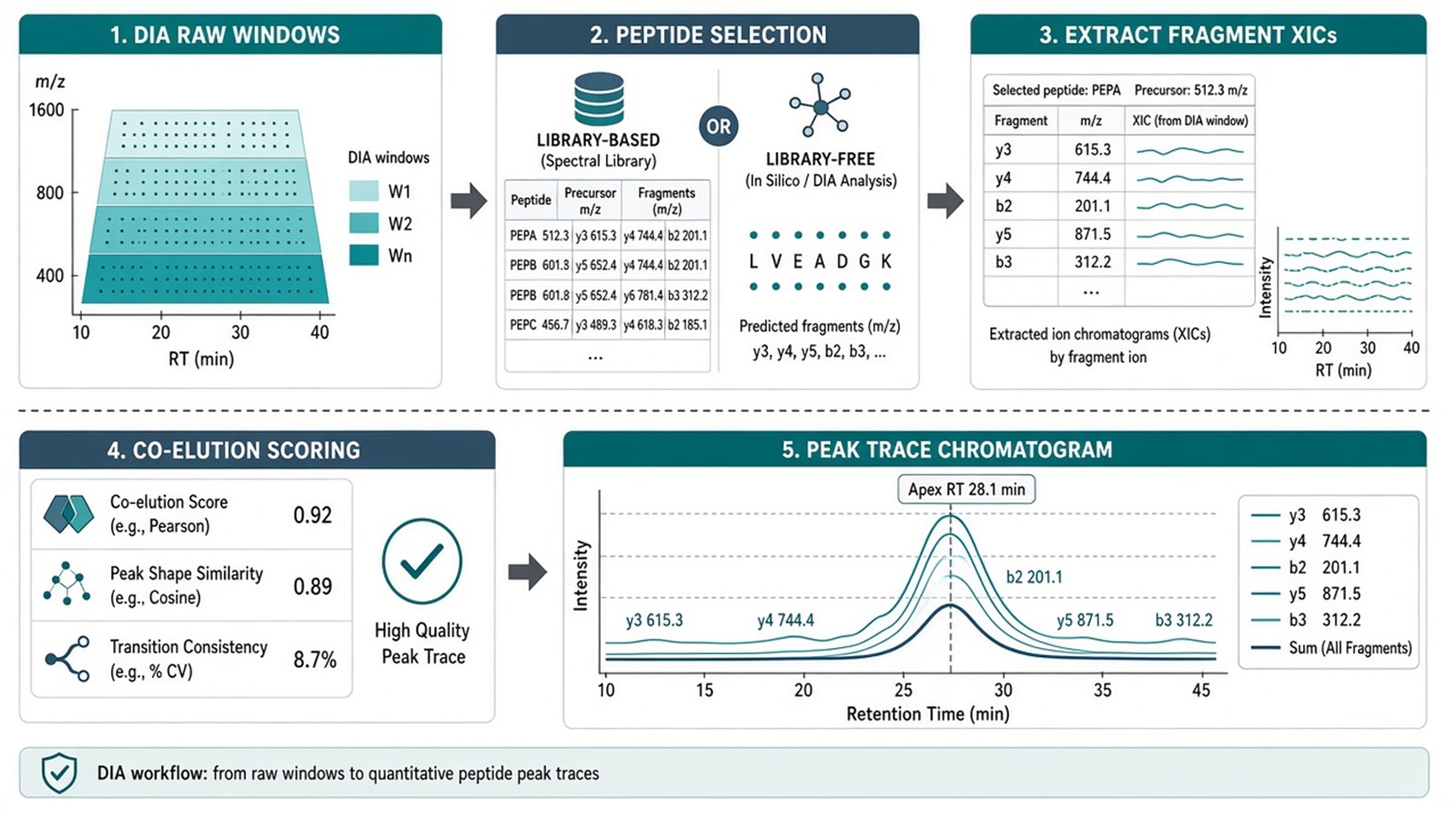
Figure 1. A specific-peptide peak trace in DIA is an FDR-filtered peak group of co-eluting fragment chromatograms, not a single raw spectrum snapshot.
A smooth chromatogram without library evidence, co-elution, and interference checks is not yet a defended peptide peak trace.
Common tools for this path include DIA-NN, Spectronaut, Skyline, and related platforms. Tool choice follows the extraction route; it does not replace matched runs or QC.
Library-Based SWATH Extraction Versus Library-Free DIA
DIA names the acquisition schedule. SWATH-MS is a sequential-window DIA implementation that historically emphasizes library-guided targeted extraction (Gillet et al. 2012). Library-free or Direct DIA routes search DIA spectra against a database, predicted fragments, or an internal library under FDR control (Demichev et al. 2020).
Route |
Advantage for a named peptide |
Matching limit |
|---|---|---|
Library-based SWATH-style |
Clear transition list and RT priors when the library matches the matrix |
Wrong or incomplete library entries yield missing or false traces |
Library-free / predicted DIA |
No separate DDA library block for that project |
Still carries window interference and FDR burden |
Manual XIC in viewer software |
Useful for visual QC of one peptide |
Not scalable and easy to over-interpret noise |
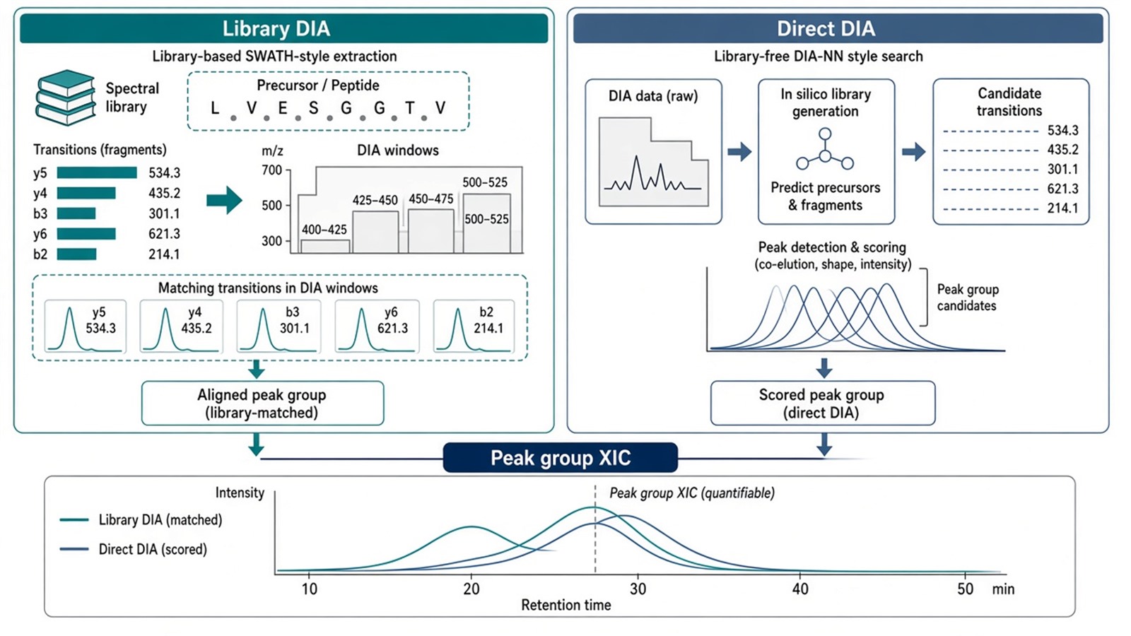
Figure 2. Library-based and library-free DIA both aim at peptide peak groups; they differ in how fragment expectations are supplied, not in the need for co-elution QC.
For DIA versus SWATH framing, see What Is DIA and SWATH? Understanding Data-Independent Acquisition in Proteomics and What Is SWATH Mass Spectrometry?. For library-free design, see Direct DIA: High-Efficiency Proteomics Without a Spectral Library. For tool pairing, see Essential Analysis Tools for Label-Free Proteomics: A Comprehensive Review of MaxQuant and DIA-NN.
Judgment Criteria: When DIA Peak-Trace Analysis Fits
Study need |
DIA or SWATH peak-trace analysis often fits |
Reassess before trusting the XIC |
|---|---|---|
Question |
Relative chromatogram for a known or discoverable peptide |
Absolute concentration or diagnostic cutoff |
Acquisition |
Matched DIA window scheme across compared runs |
Mixed window maps or unstable chromatography |
Evidence |
Library entry or library-free hit passes FDR |
Peptide absent from library and search space |
Interference |
Co-eluting transitions agree in shape and timing |
Chimeric fragments or split peaks dominate |
Panel status |
Discovery or mid-size relative follow-up |
Closed shortlist needing PRM or MRM ion control |
Prefer DIA peak-trace extraction when runs share one DIA plan and the peptide can be defended as a peak group. Prefer SWATH-style library extraction when a matrix-matched library already exists. Prefer library-free DIA when building a separate DDA library is not practical. Prefer PRM or MRM when a short closed list needs tighter precursor selection. Prefer AQUA or other SIS assays when amount units are required.
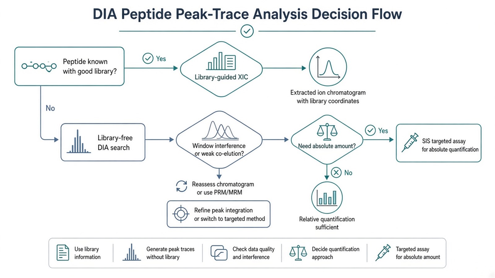
Figure 3. Choose the DIA extraction route from library readiness and QC; change method when interference or absolute claims exceed DIA peak-trace limits.
For DIA advantage lists, see Advantages and Disadvantages of DIA in Protein Quantification. For SWATH versus DDA selection, see SWATH-MS vs DDA-MS: Which Technique Is Suitable for Your Research?. For DIA versus DDA inside LFQ, see DIA vs. DDA in Label-Free Quantitative Proteomics: A Comparative Analysis.
Limits and Application Boundaries
A peak trace does not prove protein activity. Completeness in DIA is not accuracy. Window interference can invent plausible but wrong chromatograms if FDR and shape filters are weak. Cross-run comparison of peak areas assumes matched digestion, chromatography, and normalization; software extraction cannot repair unmatched prep. Pathway enrichment from proteins inferred from those peptides remains relative overrepresentation, not pathway proof.
Typical applications:
- Inspecting a biomarker-candidate peptide after DIA discovery.
- Confirming that a differential protein is supported by clean peptide peak groups.
- Comparing relative peak areas of the same peptide across treatment groups under one DIA scheme.
- Teaching or reporting how SWATH-style library extraction yields fragment chromatograms.
Diagnostic cutoffs and clinical decision limits sit outside ordinary research DIA peak-trace reporting unless a separately validated assay framework is in place.
Once the extraction route, FDR rules, and relative claim are defined, the project can be scoped as a DIA or SWATH analysis with peptide-level QC. Researchers can review the MtoZ Biolabs Deep DIA Proteomics Service for sample evaluation and workflow planning. For unlabeled DIA service framing, see Label-Free DIA Quantitative Proteomics.
Frequently Asked Questions
1. Is a DIA peak trace the same as a DDA MS1 peak?
No. DIA peak traces are usually fragment-ion chromatograms from scheduled windows. DDA quantification often starts from precursor MS1 features after stochastic MS2 selection.
2. Do I always need a spectral library to see a peptide peak?
No. Library-free or predicted routes can generate peak groups. They still need FDR control and interference QC.
3. Why does my peptide show a peak in one sample but not another?
Common causes are retention-time drift, missing library evidence, window interference, true biological absence, or failed peak-group scoring. Check co-elution and FDR before interpreting biology.
4. Can peak-trace area give absolute peptide concentration?
Not by itself. Absolute amount needs matched standards and calibration, typically on a closed targeted panel.
5. When should I move from DIA XIC review to PRM or MRM?
When the peptide list is closed, interference remains high, or the claim needs tighter ion control than wide DIA windows provide.
Reference
- L.C. Gillet, P. Navarro, S. Tate, et al. (2012). Targeted data extraction of the MS/MS spectra generated by data-independent acquisition: a new concept for consistent and accurate proteome analysis. Mol. Cell. Proteomics, 11, O111.016717. https://doi.org/10.1074/mcp.M111.016717
- V. Demichev, C.B. Messner, S.I. Vernardis, K.S. Lilley, M. Ralser (2020). DIA-NN: neural networks and interference correction enable deep proteome coverage in high throughput. Nat. Methods, 17, 41-44. https://doi.org/10.1038/s41592-019-0638-x
